The BNIP3 Antibody (CAB5683) is a high-quality antibody developed for reliable detection and analysis of target proteins. This antibody, produced in rabbits, exhibits high specificity and sensitivity in detecting BNIP3 in human samples, making it ideal for Western blot applications. By binding to the BNIP3 protein, this antibody enables precise detection and analysis of BNIP3 expression in various cell types, facilitating research in the fields of cell biology, cancer biology, and neurology.BNIP3, a member of the Bcl-2 family of proteins, plays a crucial role in regulating cell survival and death pathways by promoting mitochondrial dysfunction and autophagy.
This antibody is validated for use in WB, IHC-P, ELISA applications and has demonstrated reactivity against Human, Mouse, Rat samples.
Product Name:
BNIP3 Antibody
SKU:
CAB5683
Size:
20μL, 100μL
Reactivity:
Human, Mouse, Rat
Conjugate:
Unconjugated
Immunogen:
Recombinant protein (or fragment).This information is considered to be commercially sensitive.
This gene is encodes a mitochondrial protein that contains a BH3 domain and acts as a pro-apoptotic factor. The encoded protein interacts with anti-apoptotic proteins, including the E1B 19 kDa protein and Bcl2. This gene is silenced in tumors by DNA methylation.
Purification Method
Affinity purification
Gene ID
664
RRID
AB_2766443
Buffer Information
Store at -20℃. Avoid freeze / thaw cycles. Buffer: PBS containing 50% glycerol, preserved with proclin300 or sodium azide, pH 7.3.
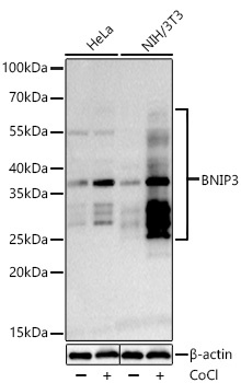
Western blot analysis of various lysates using BNIP3 Rabbit pAb (CAB5683) at 1:1000 dilution incubated overnight at 4℃. Hela and NIH/3T3 cells were treated by Cobalt chloride (0.1 mM) at 37℃ for 4 hours. Secondary antibody: HRP-conjugated Goat anti-Rabbit IgG (H+L) (CABS014) at 1:10000 dilution. Lysates/proteins: 30 μg per lane. Blocking buffer: 3% nonfat dry milk in TBST. Detection: ECL Basic Kit (AbGn00020). Exposure time: 45s.
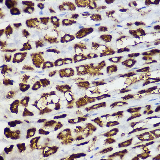
Immunohistochemistry analysis of paraffin-embedded Mouse stomach using BNIP3 Rabbit pAb (CAB5683) at dilution of 1:100 (40x lens). High pressure antigen retrieval performed with 0.01M Citrate buffer (pH 6.0) prior to IHC staining.