The BST2 Antibody (CAB12315) is a high-quality antibody developed for reliable detection and analysis of target proteins. This antibody is produced in rabbits and has been shown to be highly specific for binding to human samples, making it an ideal choice for a wide range of research applications.BST2, also known as tetherin, is a key player in the innate immune response to viral infections, particularly in restricting the release of enveloped viruses from infected cells. Understanding the role of BST2 in viral pathogenesis and immune evasion is crucial for developing strategies to combat viral infections and enhance immune defenses.
This antibody is validated for use in WB, IHC-P, IF/ICC, ELISA applications and has demonstrated reactivity against Human, Mouse samples.
Product Name:
BST2 Antibody
SKU:
CAB12315
Size:
20μL, 100μL
Reactivity:
Human, Mouse
Conjugate:
Unconjugated
Immunogen:
Recombinant protein (or fragment).This information is considered to be commercially sensitive.
Sequence:
NSEA CRDG LRAV MECR NVTH LLQQ ELTE AQKG FQDV EAQA ATCN HTVM ALMA SLDA EKAQ GQKK VEEL EGEI TTLN HKLQ DASA EVER LRRE NQVL SVRI ADKK YYPS SQDS S
Tested Applications:
WBIHC-PIF/ICCELISA
Recommended Dilution:
WB
1:500 - 1:2000
IHC-P
1:50 - 1:200
IF/ICC
1:50 - 1:200
ELISA
Recommended starting concentration is 1 μg/mL. Please optimize the concentration based on your specific assay requirements.
Synonyms:
CD317, HM1.24, TETHERIN, BST2
Positive Sample:
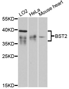
LO2, HeLa, Mouse heart
Cellular Localization:
Apical Cell Membrane, Cell Membrane, Cytoplasm, Gpi-Anchor, Golgi Apparatus, Late Endosome, Lipid-Anchor, Membrane Raft, Single-Pass Type Ii Membrane Protein, Trans-Golgi Network.
Calculated MW:
20kDa
Observed MW:
35-40kDa
Bone marrow stromal cells are involved in the growth and development of B-cells. The specific function of the protein encoded by the bone marrow stromal cell antigen 2 is undetermined; however, this protein may play a role in pre-B-cell growth and in rheumatoid arthritis.
Purification Method
Affinity purification
Gene ID
684
RRID
AB_2759169
Buffer Information
Store at -20℃. Avoid freeze / thaw cycles. Buffer: Buffer: PBS containing 50% glycerol, preserved with proclin300 or sodium azide, pH 7.3.
Western blot analysis of various lysates using BST2 Rabbit pAb (CAB12315) at 1:3000 dilution. Secondary antibody: HRP-conjugated Goat anti-Rabbit IgG (H+L) (CABS014) at 1:10000 dilution. Lysates/proteins: 25μg per lane. Blocking buffer: 3% nonfat dry milk in TBST. Detection: ECL Basic Kit (AbGn00020). Exposure time: 90s.
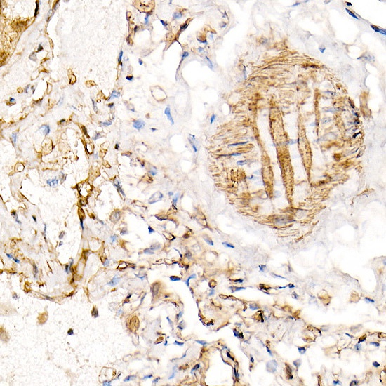
Immunohistochemistry analysis of paraffin-embedded Human lung using BST2 Rabbit pAb (CAB12315) at dilution of 1:100 (40x lens). High pressure antigen retrieval performed with 0.01M Citrate buffer (pH 6.0) prior to IHC staining.
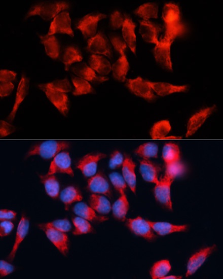
Immunofluorescence analysis of HeLa cells using BST2 Rabbit pAb (CAB12315) at dilution of 1:100. Secondary antibody: Cy3-conjugated Goat anti-Rabbit IgG (H+L) (CABS007) at 1:500 dilution. Blue: DAPI for nuclear staining.