The C11orf30 Antibody (CAB6138) is a high-quality antibody developed for reliable detection and analysis of target proteins. This antibody, produced in rabbits, exhibits high specificity and reactivity with human samples, making it ideal for use in Western blot applications. By targeting the C11orf30 protein, this antibody enables researchers to detect and analyze its expression in a variety of cell types.C11orf30 is a protein whose function is still being elucidated, with some studies suggesting a role in cellular metabolism and signaling pathways. Research into C11orf30 is particularly relevant in the fields of cell biology and cancer research, where understanding its function may provide insights into disease mechanisms and potential therapeutic targets.
This antibody is validated for use in WB, IF/ICC, ELISA applications and has demonstrated reactivity against Human, Mouse samples.
Product Name:
C11orf30 Antibody
SKU:
CAB6138
Size:
20μL, 100μL
Reactivity:
Human, Mouse
Conjugate:
Unconjugated
Immunogen:
Recombinant protein (or fragment).This information is considered to be commercially sensitive.
Recommended starting concentration is 1 μg/mL. Please optimize the concentration based on your specific assay requirements.
Synonyms:
GL002, C11orf30
Positive Sample:
MCF-7
Cellular Localization:
Nucleus.
Calculated MW:
141kDa
Observed MW:
141kDa
Predicted to enable identical protein binding activity. Predicted to be involved in DNA repair; chromatin organization; and regulation of transcription, DNA-templated. Located in nucleoplasm.
Purification Method
Affinity purification
Gene ID
56946
RRID
AB_2766768
Buffer Information
Store at -20℃. Avoid freeze / thaw cycles. Buffer: PBS containing 50% glycerol, preserved with proclin300 or sodium azide, pH 7.3.
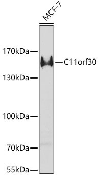
Western blot analysis of lysates from MCF-7 cells, using C11orf30 Rabbit pAb (CAB6138) at 1:1000 dilution. Secondary antibody: HRP-conjugated Goat anti-Rabbit IgG (H+L) (CABS014) at 1:10000 dilution. Lysates/proteins: 25μg per lane. Blocking buffer: 3% nonfat dry milk in TBST. Detection: ECL Enhanced Kit (AbGn00021). Exposure time: 60s.
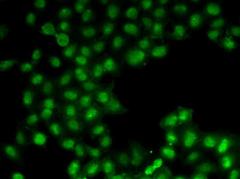
Immunofluorescence analysis of U2OS cells using C11orf30 Rabbit pAb (CAB6138).Secondary antibody: Cy3-conjugated Goat anti-Rabbit IgG (H+L) (CABS007) at 1:500 dilution.