The C7 Antibody (CAB5394) is a high-quality antibody developed for reliable detection and analysis of target proteins. This antibody, generated in rabbits, exhibits high reactivity with human samples and has been validated for use in Western blot applications. By specifically binding to the C7 protein, this antibody allows for the detection and analysis of C7 in a variety of cell types.C7 is known for its involvement in immune function and has been linked to various diseases, including cancer and autoimmune disorders.
This antibody is validated for use in WB, IF/ICC, ELISA applications and has demonstrated reactivity against Human, Mouse, Rat samples.
Product Name:
C7 Antibody
SKU:
CAB5394
Size:
20μL, 100μL
Reactivity:
Human, Mouse, Rat
Conjugate:
Unconjugated
Immunogen:
Synthetic peptide. This information is considered to be commercially sensitive.
Recommended starting concentration is 1 μg/mL. Please optimize the concentration based on your specific assay requirements.
Synonyms:
C7
Positive Sample:
Mouse plasma, Rat plasma
Cellular Localization:
Secreted.
Calculated MW:
94kDa
Observed MW:
100-105kDa
This gene encodes a serum glycoprotein that forms a membrane attack complex together with complement components C5b, C6, C8, and C9 as part of the terminal complement pathway of the innate immune system. The protein encoded by this gene contains a cholesterol-dependent cytolysin/membrane attack complex/perforin-like (CDC/MACPF) domain and belongs to a large family of structurally related molecules that form pores involved in host immunity and bacterial pathogenesis. This protein initiates membrane attack complex formation by binding the C5b-C6 subcomplex and inserts into the phospholipid bilayer, serving as a membrane anchor. Mutations in this gene are associated with a rare disorder called C7 deficiency.
Purification Method
Affinity purification
Gene ID
730
RRID
AB_2766203
Buffer Information
Store at -20℃. Avoid freeze / thaw cycles. Buffer: PBS containing 50% glycerol, preserved with proclin300 or sodium azide, pH 7.3.
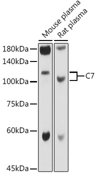
Western blot analysis of various lysates using C7 Rabbit pAb (CAB5394) at 1:1000 dilution. Secondary antibody: HRP-conjugated Goat anti-Rabbit IgG (H+L) (CABS014) at 1:10000 dilution. Lysates/proteins: 25μg per lane. Blocking buffer: 3% nonfat dry milk in TBST. Detection: ECL Basic Kit (AbGn00020). Exposure time: 1s.
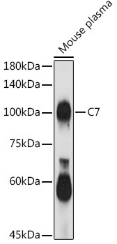
Western blot analysis of lysates from Mouse plasma, using C7 Rabbit pAb (CAB5394) at 1:1000 dilution. Secondary antibody: HRP-conjugated Goat anti-Rabbit IgG (H+L) (CABS014) at 1:10000 dilution. Lysates/proteins: 25μg per lane. Blocking buffer: 3% nonfat dry milk in TBST. Detection: ECL Basic Kit (AbGn00020). Exposure time: 180s.
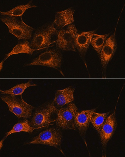
Immunofluorescence analysis of NIH/3T3 cells using C7 Rabbit pAb (CAB5394) at dilution of 1:100. Secondary antibody: Cy3-conjugated Goat anti-Rabbit IgG (H+L) (CABS007) at 1:500 dilution. Blue: DAPI for nuclear staining.