The CABP7 Antibody (CAB13703) is a high-quality antibody developed for reliable detection and analysis of target proteins. This antibody, produced in rabbits, shows high reactivity with human samples and is validated for use in various applications, including Western blot and immunohistochemistry.CABP7 is known to play a role in regulating calcium levels within cells, making it essential for processes such as neurotransmitter release and muscle contraction. Dysregulation of calcium signaling can lead to diseases such as Alzheimer's, Parkinson's, and heart conditions, making CABP7 an important target for therapeutic interventions.
This antibody is validated for use in WB, IHC-P, ELISA applications and has demonstrated reactivity against Human, Mouse, Rat samples.
Product Name:
CABP7 Antibody
SKU:
CAB13703
Size:
20μL, 100μL
Reactivity:
Human, Mouse, Rat
Conjugate:
Unconjugated
Immunogen:
Recombinant protein (or fragment).This information is considered to be commercially sensitive.
Recommended starting concentration is 1 μg/mL. Please optimize the concentration based on your specific assay requirements.
Synonyms:
CALN2, CABP7
Positive Sample:
Mouse brain, Mouse lung, Mouse kidney, Rat lung, Rat kidney
Cellular Localization:
Cell Membrane, Cytoplasm, Golgi Apparatus, Single-Pass Type Iv Membrane Protein, Perinuclear Region, Trans-Golgi Network Membrane.
Calculated MW:
24kDa
Observed MW:
30kDa
Predicted to enable calcium ion binding activity. Located in trans-Golgi network membrane.
Purification Method
Affinity purification
Gene ID
164633
RRID
AB_2760564
Buffer Information
Store at -20℃. Avoid freeze / thaw cycles. Buffer: PBS with 0.01% thimerosal,50% glycerol,pH7.3.
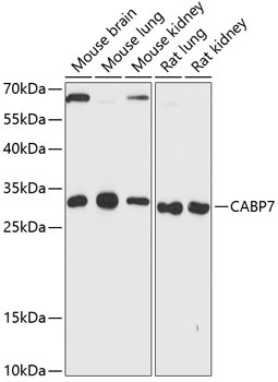
Western blot analysis of various lysates using CABP7 Rabbit pAb (CAB13703) at 1:3000 dilution. Secondary antibody: HRP-conjugated Goat anti-Rabbit IgG (H+L) (CABS014) at 1:10000 dilution. Lysates/proteins: 25μg per lane. Blocking buffer: 3% nonfat dry milk in TBST. Detection: ECL Enhanced Kit (AbGn00021). Exposure time: 90s.
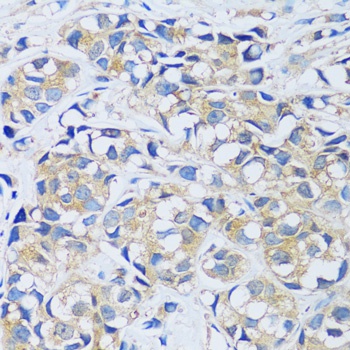
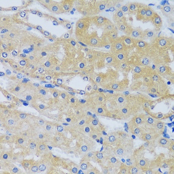
Immunohistochemistry analysis of paraffin-embedded Human breast cancer using CABP7 Rabbit pAb (CAB13703) at dilution of 1:100 (40x lens). Microwave antigen retrieval performed with 0.01M PBS Buffer (pH 7.2) prior to IHC staining.
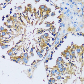
Immunohistochemistry analysis of paraffin-embedded Mouse testis using CABP7 Rabbit pAb (CAB13703) at dilution of 1:100 (40x lens). Microwave antigen retrieval performed with 0.01M PBS Buffer (pH 7.2) prior to IHC staining.
Western blot analysis of various lysates using CABP7 Rabbit pAb (CAB13703) at 1:3000 dilution. Secondary antibody: HRP-conjugated Goat anti-Rabbit IgG (H+L) (CABS014) at 1:10000 dilution. Lysates/proteins: 25μg per lane. Blocking buffer: 3% nonfat dry milk in TBST. Detection: ECL Enhanced Kit (AbGn00021). Exposure time: 90s.