The CAD Antibody (CAB8344) is a high-quality antibody developed for reliable detection and analysis of target proteins. The antibody, produced in rabbits, shows high reactivity with CAD protein in human samples and is validated for use in Western blotting applications. By targeting the CAD protein, this antibody allows for precise detection and analysis in various cell types, making it an invaluable asset for research in molecular biology and drug development.CAD, or carbamoyl-phosphate synthetase 2, aspartate transcarbamylase, and dihydroorotase, is essential for the de novo synthesis of pyrimidines, which are building blocks for DNA and RNA.
This antibody is validated for use in WB, IHC-P, IF/ICC, ELISA applications and has demonstrated reactivity against Human, Mouse, Rat samples.
Product Name:
CAD Antibody
SKU:
CAB8344
Size:
20μL, 100μL
Reactivity:
Human, Mouse, Rat
Conjugate:
Unconjugated
Immunogen:
Recombinant protein (or fragment).This information is considered to be commercially sensitive.
Recommended starting concentration is 1 μg/mL. Please optimize the concentration based on your specific assay requirements.
Synonyms:
CDG1Z, DEE50, GATD4, EIEE50, CAD
Positive Sample:
SW620, HepG2, HeLa, HL-60
Cellular Localization:
Cytoplasm, Nucleus.
Calculated MW:
243kDa
Observed MW:
243kDa
The de novo synthesis of pyrimidine nucleotides is required for mammalian cells to proliferate. This gene encodes a trifunctional protein which is associated with the enzymatic activities of the first 3 enzymes in the 6-step pathway of pyrimidine biosynthesis: carbamoylphosphate synthetase (CPS II), aspartate transcarbamoylase, and dihydroorotase. This protein is regulated by the mitogen-activated protein kinase (MAPK) cascade, which indicates a direct link between activation of the MAPK cascade and de novo biosynthesis of pyrimidine nucleotides. Alternative splicing results in multiple transcript variants encoding different isoforms.
Purification Method
Affinity purification
Gene ID
790
RRID
AB_2768677
Buffer Information
Store at -20℃. Avoid freeze / thaw cycles. Buffer: PBS containing 50% glycerol, preserved with proclin300 or sodium azide, pH 7.3.
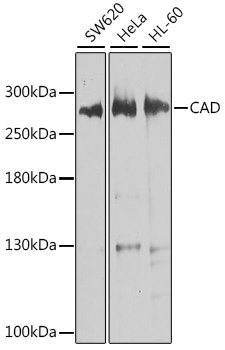
Western blot analysis of various lysates using CAD Rabbit pAb (CAB8344) at 1:1000 dilution. Secondary antibody: HRP-conjugated Goat anti-Rabbit IgG (H+L) (CABS014) at 1:10000 dilution. Lysates/proteins: 25μg per lane. Blocking buffer: 3% nonfat dry milk in TBST. Detection: ECL Basic Kit (AbGn00020). Exposure time: 10s.
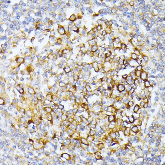
Immunohistochemistry analysis of paraffin-embedded Human tonsil using CAD Rabbit pAb (CAB8344) at dilution of 1:100 (40x lens). Microwave antigen retrieval performed with 0.01M PBS Buffer (pH 7.2) prior to IHC staining.
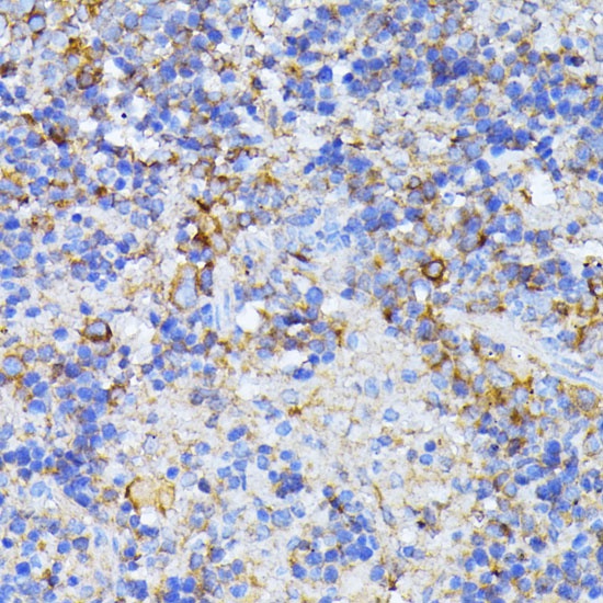
Immunohistochemistry analysis of paraffin-embedded Mouse spleen using CAD Rabbit pAb (CAB8344) at dilution of 1:100 (40x lens). Microwave antigen retrieval performed with 0.01M PBS Buffer (pH 7.2) prior to IHC staining.
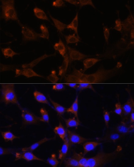
Immunofluorescence analysis of NIH-3T3 cells using CAD Rabbit pAb (CAB8344) at dilution of 1:100. Secondary antibody: Cy3-conjugated Goat anti-Rabbit IgG (H+L) (CABS007) at 1:500 dilution. Blue: DAPI for nuclear staining.