The Cadherin 16 Antibody (CAB7882) is a high-quality antibody developed for reliable detection and analysis of target proteins. This antibody, produced through rabbit immunization, shows high reactivity with human samples and is validated for use in Western blot applications. It specifically binds to the Cadherin-16 protein, enabling accurate detection and analysis in a variety of cell types, making it ideal for studies in developmental biology and cancer research.
This antibody is validated for use in WB, IHC-P, ELISA, IF-P applications and has demonstrated reactivity against Human, Mouse, Rat samples.
Product Name:
Cadherin 16 Antibody
SKU:
CAB7882
Size:
20μL, 100μL
Reactivity:
Human, Mouse, Rat
Conjugate:
Unconjugated
Immunogen:
Recombinant protein (or fragment).This information is considered to be commercially sensitive.
Recommended starting concentration is 1 μg/mL. Please optimize the concentration based on your specific assay requirements.
Synonyms:
CDH16, Cadherin 16
Positive Sample:
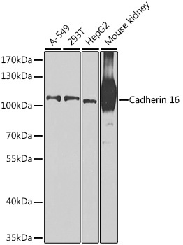
A-549, 293T, HepG2, Mouse kidney
Cellular Localization:
Cell Membrane, Single-Pass Type I Membrane Protein.
Calculated MW:
90kDa
Observed MW:
110kDa
This gene is a member of the cadherin superfamily, genes encoding calcium-dependent, membrane-associated glycoproteins. Mapped to a previously identified cluster of cadherin genes on chromosome 16q22.1, the gene localizes with superfamily members CDH1, CDH3, CDH5, CDH8 and CDH11. The protein consists of an extracellular domain containing 6 cadherin domains, a transmembrane region and a truncated cytoplasmic domain but lacks the prosequence and tripeptide HAV adhesion recognition sequence typical of most classical cadherins. Expression is exclusively in kidney, where the protein functions as the principal mediator of homotypic cellular recognition, playing a role in the morphogenic direction of tissue development. Alternatively spliced transcript variants encoding distinct isoforms have been identified.
Purification Method
Affinity purification
Gene ID
1014
RRID
AB_2768815
Buffer Information
Store at -20℃. Avoid freeze / thaw cycles. Buffer: PBS containing 50% glycerol, preserved with proclin300 or sodium azide, pH 7.3.
Western blot analysis of various lysates using Cadherin 16 Rabbit pAb (CAB7882) at 1:1000 dilution. Secondary antibody: HRP-conjugated Goat anti-Rabbit IgG (H+L) (CABS014) at 1:10000 dilution. Lysates/proteins: 25μg per lane. Blocking buffer: 3% nonfat dry milk in TBST. Detection: ECL Basic Kit (AbGn00020). Exposure time: 90s.
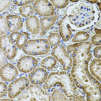
Immunohistochemistry analysis of paraffin-embedded Rat kidney using Cadherin 16 Rabbit pAb (CAB7882) at dilution of 1:100 (40x lens). Microwave antigen retrieval performed with 0.01M PBS Buffer (pH 7.2) prior to IHC staining.
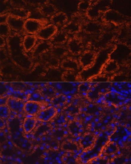
Immunofluorescence analysis of paraffin-embedded rat kidney using Cadherin 16 Rabbit pAb (CAB7882) at dilution of 1:100 (40x lens). Secondary antibody: Cy3-conjugated Goat anti-Rabbit IgG (H+L) (CABS007) at 1:500 dilution. Blue: DAPI for nuclear staining.