The CALD1 Antibody (CAB5366) is a high-quality antibody developed for reliable detection and analysis of target proteins. This gene encodes a calmodulin- and actin-binding protein that plays an essential role in the regulation of smooth muscle and nonmuscle contraction. The conserved domain of this protein possesses the binding activities to Ca(2+)-calmodulin, actin, tropomyosin, myosin, and phospholipids. This protein is a potent inhibitor of the actin-tropomyosin activated myosin MgATPase, and serves as a mediating factor for Ca(2+)-dependent inhibition of smooth muscle contraction. Alternative splicing of this gene results in multiple transcript variants encoding distinct isoforms.
This antibody is validated for use in WB, ELISA applications and has demonstrated reactivity against Human samples.
Product Name:
CALD1 Antibody
SKU:
CAB5366
Size:
100μL, 20μL
Reactivity:
Human
Conjugate:
Unconjugated
Immunogen:
Recombinant protein (or fragment).This information is considered to be commercially sensitive.
Tested Applications:
WBELISA
Recommended Dilution:
WB
1:500 - 1:2000
ELISA
Recommended starting concentration is 1 μg/mL. Please optimize the concentration based on your specific assay requirements.
Synonyms:
CDM, HCAD, LCAD, h-CD, H-CAD, L-CAD, NAG22, CALD1
Positive Sample:
A-431, 22Rv1, U-251 MG, U-87 MG
Cellular Localization:
Cytoplasm, Cytoskeleton, Myofibril.
Calculated MW:
93kDa
Observed MW:
93kDa/70-80kDa
This gene encodes a calmodulin- and actin-binding protein that plays an essential role in the regulation of smooth muscle and nonmuscle contraction. The conserved domain of this protein possesses the binding activities to Ca(2+)-calmodulin, actin, tropomyosin, myosin, and phospholipids. This protein is a potent inhibitor of the actin-tropomyosin activated myosin MgATPase, and serves as a mediating factor for Ca(2+)-dependent inhibition of smooth muscle contraction. Alternative splicing of this gene results in multiple transcript variants encoding distinct isoforms.
Purification Method
Affinity purification
Gene ID
800
RRID
AB_2766176
Buffer Information
Store at -20℃. Avoid freeze / thaw cycles. Buffer: PBS containing 50% glycerol, preserved with proclin300 or sodium azide, pH 7.3.
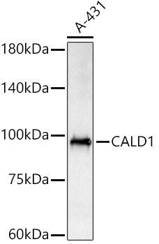
Western blot analysis of lysates from A-431 cells, using CALD1 Rabbit pAb (CAB5366) at 1:2000 dilution. Secondary antibody: HRP-conjugated Goat anti-Rabbit IgG (H+L) (AS014) at 1:10000 dilution. Lysates/proteins: 25μg per lane. Blocking buffer: 3% nonfat dry milk in TBST. Detection: ECL Basic Kit (AbGn00020). Exposure time: 180s.
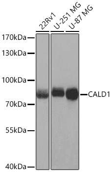
Western blot analysis of various lysates using CALD1 Rabbit pAb (CAB5366) at 1:1000 dilution incubated overnight at 4℃. Secondary antibody: HRP-conjugated Goat anti-Rabbit IgG (H+L) (AS014) at 1:10000 dilution. Lysates/proteins: 25 μg per lane. Blocking buffer: 3% nonfat dry milk in TBST. Detection: ECL Basic Kit (AbGn00020) Exposure time: 10 s.