The CaMKII delta Monoclonal Antibody (CAB9196) is a high-quality antibody developed for reliable detection and analysis of target proteins. This monoclonal antibody, generated in rabbits, is highly specific to human samples and has been validated for use in Western blot and immunohistochemistry applications.CAMKII Delta is a key player in signal transduction pathways involved in synaptic plasticity, learning, and memory. Dysregulation of CAMKII Delta has been implicated in various neurological disorders, making it a promising target for drug development and therapeutic interventions.
This antibody is validated for use in WB, IF/ICC, ELISA applications and has demonstrated reactivity against Human, Mouse samples.
Product Name:
CaMKII delta Monoclonal Antibody
SKU:
CAB9196
Size:
20μL, 100μL
Reactivity:
Human, Mouse
Clone Number:
ARC1473
Conjugate:
Unconjugated
Immunogen:
Synthetic peptide. This information is considered to be commercially sensitive.
The product of this gene belongs to the serine/threonine protein kinase family and to the Ca(2+)/calmodulin-dependent protein kinase subfamily. Calcium signaling is crucial for several aspects of plasticity at glutamatergic synapses. In mammalian cells, the enzyme is composed of four different chains: alpha, beta, gamma, and delta. The product of this gene is a delta chain. Alternative splicing results in multiple transcript variants encoding distinct isoforms. Distinct isoforms of this chain have different expression patterns.
Purification Method
Affinity purification
Gene ID
817
RRID
AB_2863685
Buffer Information
Store at -20℃. Avoid freeze / thaw cycles. Buffer: PBS containing 50% glycerol and 0.05% BSA, preserved with proclin300 or sodium azide, pH 7.3.
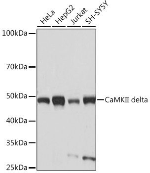
Western blot analysis of various lysates using CaMKII delta Rabbit mAb (CAB9196) at 1:1000 dilution. Secondary antibody: HRP-conjugated Goat anti-Rabbit IgG (H+L) (CABS014) at 1:10000 dilution. Lysates/proteins: 25μg per lane. Blocking buffer: 3% nonfat dry milk in TBST. Detection: ECL Basic Kit (AbGn00020). Exposure time: 10s.
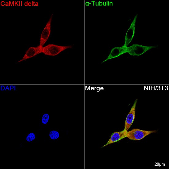
Confocal imaging of NIH/3T3 cells using CaMKII delta Rabbit mAb (CAB9196, dilution 1:100) ollowed by a further incubation with Cy3 Goat Anti-Rabbit IgG (H+L) (CABS007,dilution 1:500) (Red). The cells were counterstained with α-Tubulin Mouse mAb (AC012, dilution 1:400) followed by incubation with ABflo® 488-conjugated Goat Anti-Mouse IgG (H+L) Ab (CABS076, dilution 1:500) (Green). DAPI was used for nuclear staining (Blue). Objective: 100x.