The Calsequestrin 1 Monoclonal Antibody (CAB19640) is a high-quality antibody developed for reliable detection and analysis of target proteins. Calsequestrin-1 is a calcium-binding protein found in the sarcoplasmic reticulum of muscle cells, where it plays a crucial role in storing and releasing calcium ions during muscle contraction. This polyclonal antibody, generated in rabbits, is highly specific to human calsequestrin-1 and has been validated for use in Western blot applications.By binding to calsequestrin-1, this antibody allows for the detection and analysis of this important regulatory protein in various cell types.
This antibody is validated for use in WB, ELISA, IF-P applications and has demonstrated reactivity against Mouse, Rat samples.
Product Name:
Calsequestrin 1 Monoclonal Antibody
SKU:
CAB19640
Size:
20μL, 100μL
Reactivity:
Mouse, Rat
Clone Number:
ARC2209
Conjugate:
Unconjugated
Immunogen:
Recombinant protein (or fragment).This information is considered to be commercially sensitive.
Recommended starting concentration is 1 μg/mL. Please optimize the concentration based on your specific assay requirements.
Synonyms:
CASQ, CSQ1, PDIB1, VMCQA, Calsequestrin 1
Positive Sample:
Mouse skeletal muscle, Rat heart
Cellular Localization:
Endoplasmic Reticulum, Mitochondrial Matrix.
Calculated MW:
45kDa
Observed MW:
55kDa
This gene encodes the skeletal muscle specific member of the calsequestrin protein family. Calsequestrin functions as a luminal sarcoplasmic reticulum calcium sensor in both cardiac and skeletal muscle cells. This protein, also known as calmitine, functions as a calcium regulator in the mitochondria of skeletal muscle. This protein is absent in patients with Duchenne and Becker types of muscular dystrophy.
Purification Method
Affinity purification
Gene ID
844
Buffer Information
Store at -20℃. Avoid freeze / thaw cycles. Buffer: PBS containing 50% glycerol and 0.05% BSA, preserved with proclin300 or sodium azide, pH 7.3.
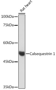
Western blot analysis of lysates from Rat heart, using Calsequestrin 1 Rabbit mAb (CAB19640) at 1:500 dilution. Secondary antibody: HRP-conjugated Goat anti-Rabbit IgG (H+L) (CABS014) at 1:10000 dilution. Lysates/proteins: 25μg per lane. Blocking buffer: 3% nonfat dry milk in TBST. Detection: ECL Basic Kit (AbGn00020). Exposure time: 60s.
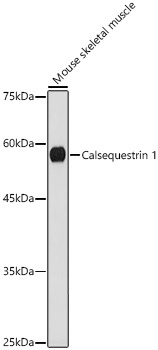
Western blot analysis of lysates from Mouse skeletal muscle, using Calsequestrin 1 Rabbit mAb (CAB19640) at 1:1000 dilution. Secondary antibody: HRP-conjugated Goat anti-Rabbit IgG (H+L) (CABS014) at 1:10000 dilution. Lysates/proteins: 25μg per lane. Blocking buffer: 3% nonfat dry milk in TBST. Detection: ECL Basic Kit (AbGn00020). Exposure time: 180s.
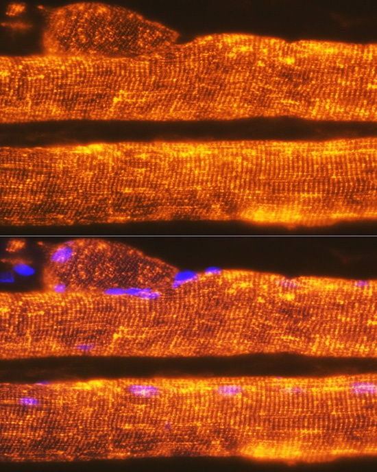
Immunofluorescence analysis of paraffin-embedded Rat skeletal muscle using Calsequestrin 1 Rabbit mAb (CAB19640) at dilution of 1:100. Secondary antibody: Cy3-conjugated Goat anti-Rabbit IgG (H+L) (CABS007) at 1:500 dilution. Blue: DAPI for nuclear staining.