The CD13 Polyclonal Antibody (CAB23653) is a high-quality antibody developed for reliable detection and analysis of target proteins. This antibody, produced in rabbits, is highly specific to human CD13 and is validated for use in Western blot applications. By binding to the CD13 protein, this antibody allows for accurate detection and analysis in a wide range of cell types, making it an essential reagent for studies in immunology, cancer research, and more.CD13, also known as Aminopeptidase N, is involved in processes such as cell adhesion, migration, and angiogenesis, making it a key player in tumor development and progression.
This antibody is validated for use in WB, IHC-P, ELISA applications and has demonstrated reactivity against Human, Mouse, Rat samples.
Product Name:
CD13 Polyclonal Antibody
SKU:
CAB23653
Size:
20μL, 100μL
Reactivity:
Human, Mouse, Rat
Conjugate:
Unconjugated
Immunogen:
Recombinant protein (or fragment).This information is considered to be commercially sensitive.
Sequence:
Email for sequence
Tested Applications:
WBIHC-PELISA
Recommended Dilution:
WB
1:500 - 1:1000
IHC-P
1:500 - 1:1000
ELISA
Recommended starting concentration is 1 μg/mL. Please optimize the concentration based on your specific assay requirements.
Synonyms:
Apn, AP-M, AP-N, Cd13, P150, CD13
Positive Sample:
HepG2, Mouse kidney, Rat kidney
Calculated MW:
109kDa
Observed MW:
160kDa
Predicted to enable metalloaminopeptidase activity; peptide binding activity; and zinc ion binding activity. Predicted to be involved in several processes, including negative regulation of renal sodium excretion; peptide catabolic process; and proteolysis. Predicted to act upstream of or within angiogenesis and cell differentiation. Located in external side of plasma membrane. Is expressed in several structures, including alimentary system; brain; metanephros; reproductive system; and sensory organ. Orthologous to human ANPEP (alanyl aminopeptidase, membrane).
Purification Method
Affinity purification
Gene ID
16790
Buffer Information
Store at -20℃. Avoid freeze / thaw cycles. Buffer: PBS containing 50% glycerol, preserved with proclin300 or sodium azide, pH 7.3.
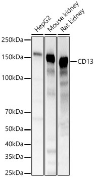
Western blot analysis of various lysates, using CD13 Rabbit pAb (CAB23653) at 1:1000 dilution. Secondary antibody: HRP-conjugated Goat anti-Rabbit IgG (H+L) (CABS014) at 1:10000 dilution. Lysates/proteins: 25μg per lane. Blocking buffer: 3% nonfat dry milk in TBST. Detection: ECL Basic Kit (AbGn00020). Exposure time: 1s.
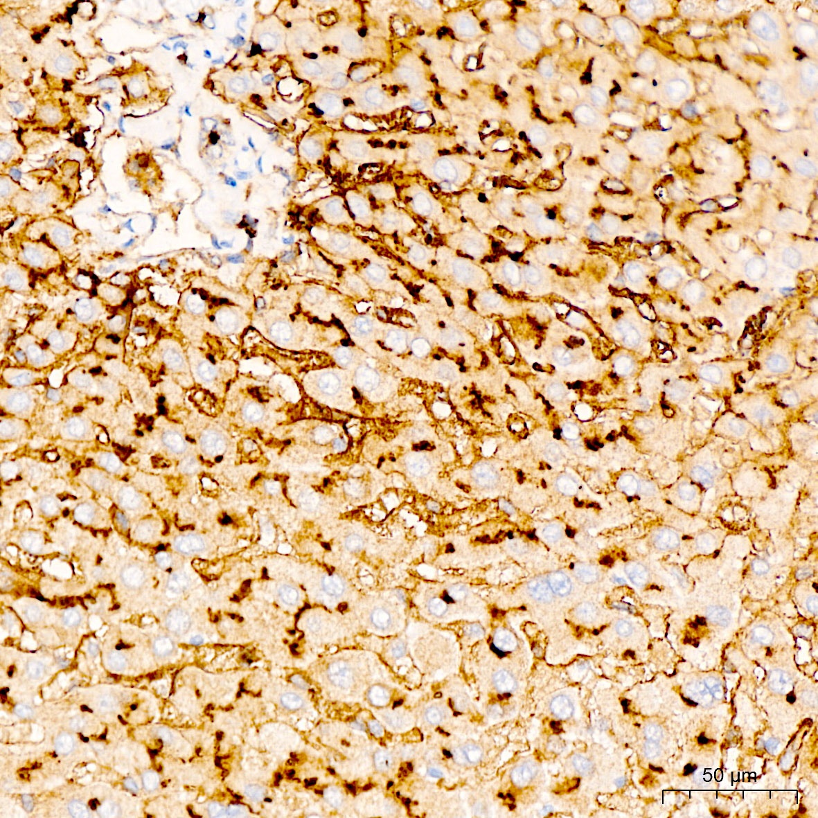
Immunohistochemistry analysis of paraffin-embedded Human liver tissue using CD13 Rabbit pAb (CAB23653) at a dilution of 1:900 (40x lens). High pressure antigen retrieval was performed with 0.01 M citrate buffer (pH 6.0) prior to IHC staining.
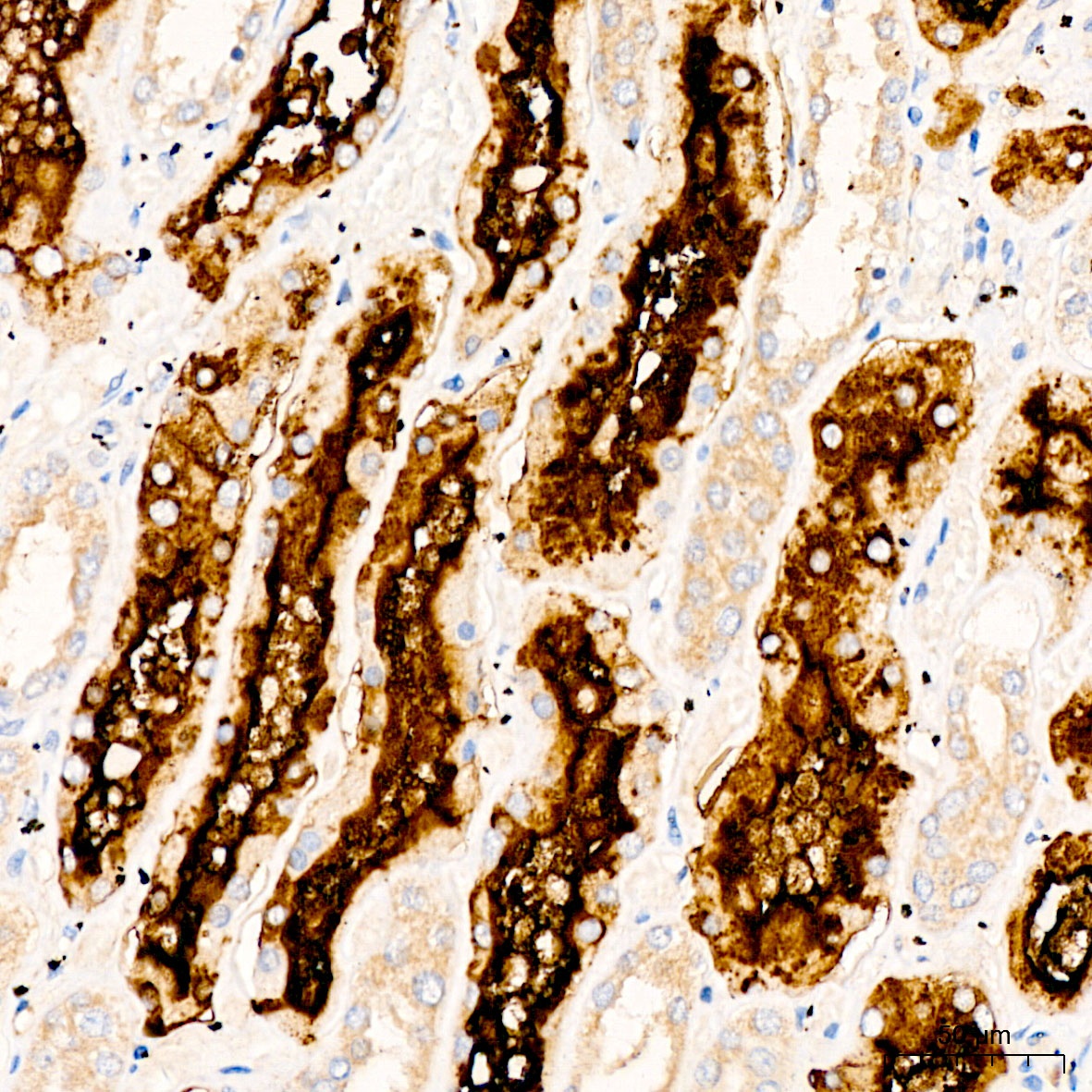
Immunohistochemistry analysis of paraffin-embedded Rat kidney tissue using CD13 Rabbit pAb (CAB23653) at a dilution of 1:900 (40x lens). High pressure antigen retrieval was performed with 0.01 M citrate buffer (pH 6.0) prior to IHC staining.
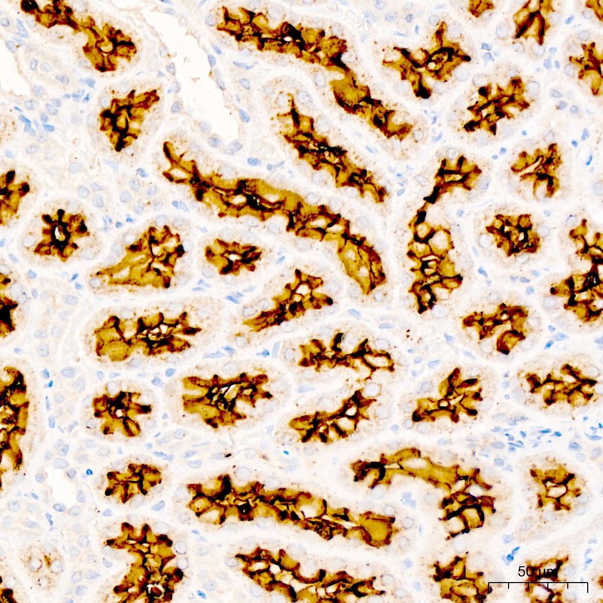
Immunohistochemistry analysis of paraffin-embedded Human kidney tissue using CD13 Rabbit pAb (CAB23653) at a dilution of 1:900 (40x lens). High pressure antigen retrieval was performed with 0.01 M citrate buffer (pH 6.0) prior to IHC staining.
 at 1:1000 dilution. Secondary antibody: HRP Goat Anti-Rabbit IgG (H+L) at 1:10000 dilution. Lysates/proteins: 25μg per lane. Blocking buffer: 3% nonfat dry milk in TBST.")
 at 1:1000 dilution. Secondary antibody: HRP Goat Anti-Rabbit IgG (H+L) at 1:10000 dilution. Lysates/proteins: 25μg per lane. Blocking buffer: 3% nonfat dry milk in TBST.")


