The CD3E Monoclonal Antibody (CAB19017) is a high-quality antibody developed for reliable detection and analysis of target proteins. The protein encoded by this gene is the CD3-epsilon polypeptide, which together with CD3-gamma, -delta and -zeta, and the T-cell receptor alpha/beta and gamma/delta heterodimers, forms the T-cell receptor-CD3 complex. This complex plays an important role in coupling antigen recognition to several intracellular signal-transduction pathways. The genes encoding the epsilon, gamma and delta polypeptides are located in the same cluster on chromosome 11. The epsilon polypeptide plays an essential role in T-cell development. Defects in this gene cause immunodeficiency. This gene has also been linked to a susceptibility to type I diabetes in women.
This antibody is validated for use in WB, IHC-P, IF/ICC, ELISA, IF-P, mIHC applications and has demonstrated reactivity against Human, Mouse, Rat samples.
Product Name:
CD3E Monoclonal Antibody
SKU:
CAB19017
Size:
100μL
Reactivity:
Human, Mouse, Rat
Clone Number:
ARC51750
Conjugate:
Unconjugated
Immunogen:
Synthetic peptide. This information is considered to be commercially sensitive.
Tested Applications:
WBIHC-PIF/ICCELISAIF-PmIHC
Recommended Dilution:
WB
1:10000 - 1:60000
IF/ICC
1:200-1:800
IF-P
1:200-1:800
IHC-P
1:1000 - 1:5000
mIHC
1:1000 - 1:5000
ELISA
Recommended starting concentration is 1 μg/mL. Please optimize the concentration based on your specific assay requirements.
Synonyms:
T3E, TCRE, IMD18, CD3epsilon, CD3E
Positive Sample:
Mouse spleen, Jurkat, MOLT-4
Cellular Localization:
Cell Membrane, Single-Pass Type I Membrane Protein.
Calculated MW:
23kDa
Observed MW:
23kDa
The protein encoded by this gene is the CD3-epsilon polypeptide, which together with CD3-gamma, -delta and -zeta, and the T-cell receptor alpha/beta and gamma/delta heterodimers, forms the T-cell receptor-CD3 complex. This complex plays an important role in coupling antigen recognition to several intracellular signal-transduction pathways. The genes encoding the epsilon, gamma and delta polypeptides are located in the same cluster on chromosome 11. The epsilon polypeptide plays an essential role in T-cell development. Defects in this gene cause immunodeficiency. This gene has also been linked to a susceptibility to type I diabetes in women.
Purification Method
Affinity purification
Gene ID
916
RRID
AB_2862509
Buffer Information
Store at -20℃. Avoid freeze / thaw cycles. Buffer: PBS containing 50% glycerol and 0.05% BSA, preserved with proclin300 or sodium azide, pH 7.3.
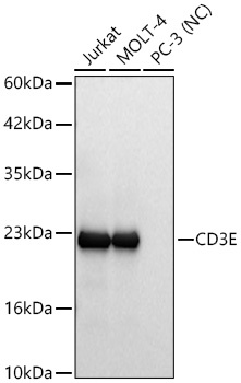
Western blot analysis of various lysates using CD3E Rabbit mAb (CAB19017) at 1:10000 dilution incubated overnight at 4℃. Secondary antibody: HRP-conjugated Goat anti-Rabbit IgG (H+L) (AS014) at 1:10000 dilution. Lysates/proteins: 25 μg per lane. Blocking buffer: 3% nonfat dry milk in TBST. Detection: ECL Basic Kit (AbGn00020). Negative control (NC): PC-3 Exposure time: 5s.
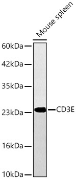
Western blot analysis of lysates from Mouse spleen using CD3E Rabbit mAb (CAB19017) at 1:10000 dilution incubated overnight at 4℃. Secondary antibody: HRP-conjugated Goat anti-Rabbit IgG (H+L) (AS014) at 1:10000 dilution. Lysates/proteins: 25 μg per lane. Blocking buffer: 3% nonfat dry milk in TBST. Detection: ECL Basic Kit (AbGn00020). Exposure time: 10s.
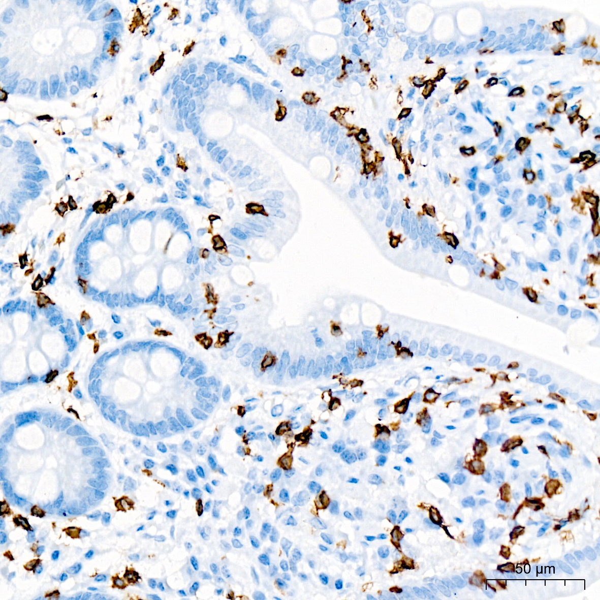
Immunohistochemistry analysis of paraffin-embedded Human small intestine tissue using CD3E Rabbit mAb (CAB19017) at a dilution of 1:2000 (40x lens). High pressure antigen retrieval was performed with 0.01 M citrate buffer (pH 6.0) prior to IHC staining.
Immunohistochemistry analysis of paraffin-embedded Rat spleen tissue using CD3E Rabbit mAb (CAB19017) at a dilution of 1:2000 (40x lens). High pressure antigen retrieval was performed with 0.01 M citrate buffer (pH 6.0) prior to IHC staining.
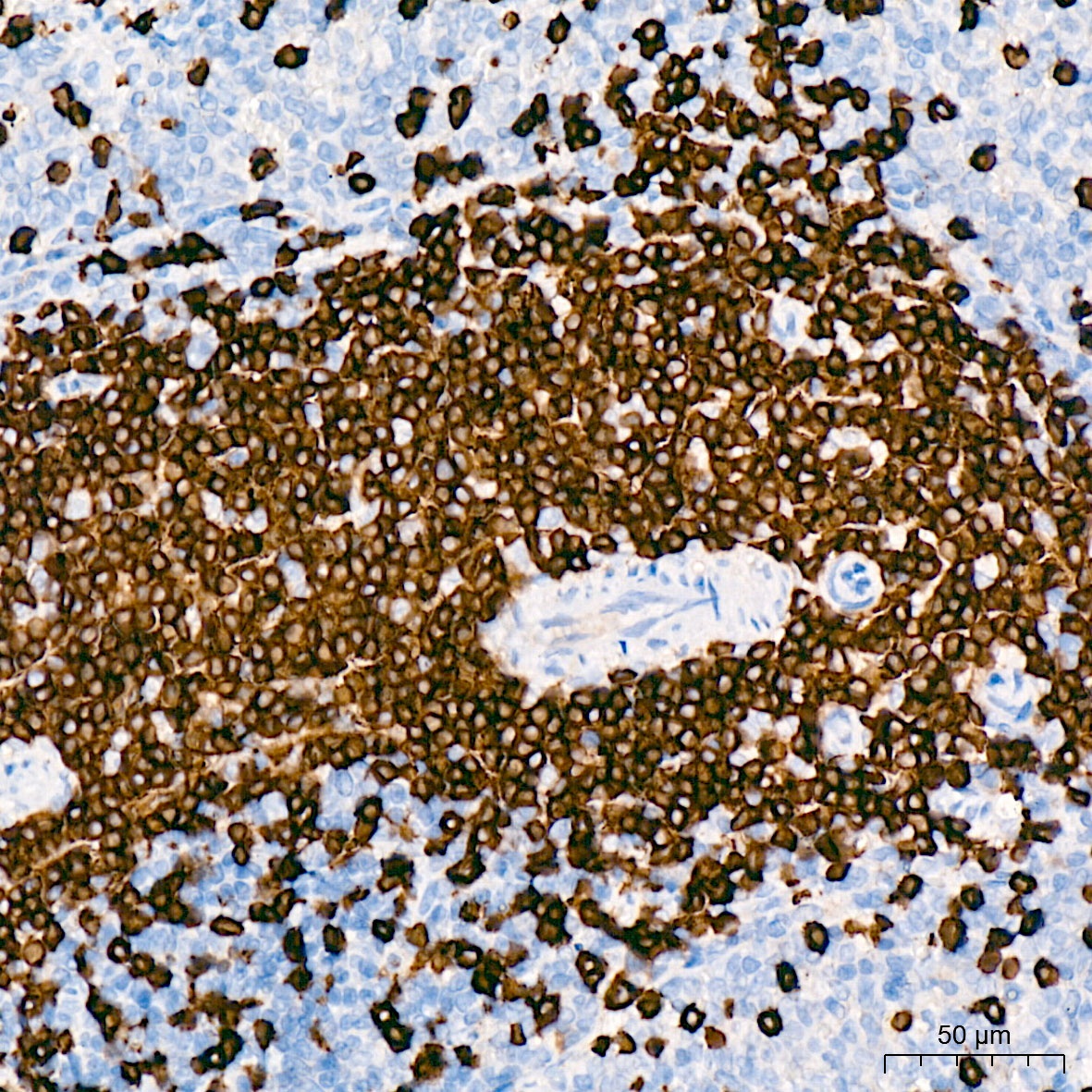
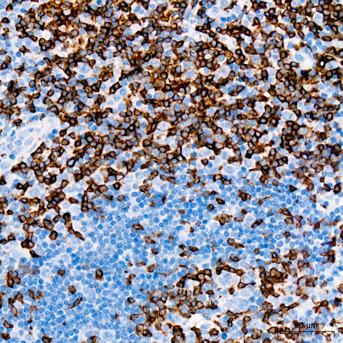
Immunohistochemistry analysis of paraffin-embedded Human tonsil tissue using CD3E Rabbit mAb (CAB19017) at a dilution of 1:2000 (40x lens). High pressure antigen retrieval was performed with 0.01 M citrate buffer (pH 6.0) prior to IHC staining.
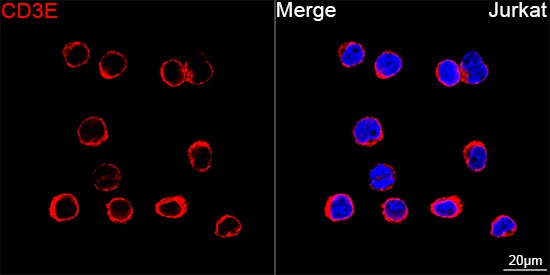
Confocal imaging of Jurkat cells using CD3E Rabbit mAb (CAB19017, dilution 1:200) followed by a further incubation with Cy3 Goat Anti-Rabbit IgG (H+L) (AS007, dilution 1:500) (Red). DAPI was used for nuclear staining (Blue). Objective: 100x.
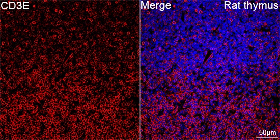
Confocal imaging of paraffin-embedded Rat thymus tissue using CD3E Rabbit mAb (CAB19017, dilution 1:200) followed by a further incubation with Cy3 Goat Anti-Rabbit IgG (H+L) (AS007, dilution 1:500) (Red). DAPI was used for nuclear staining (Blue). High pressure antigen retrieval performed with 0.01M Citrate Buffer (pH 6.0) prior to IF staining. Objective: 40x.
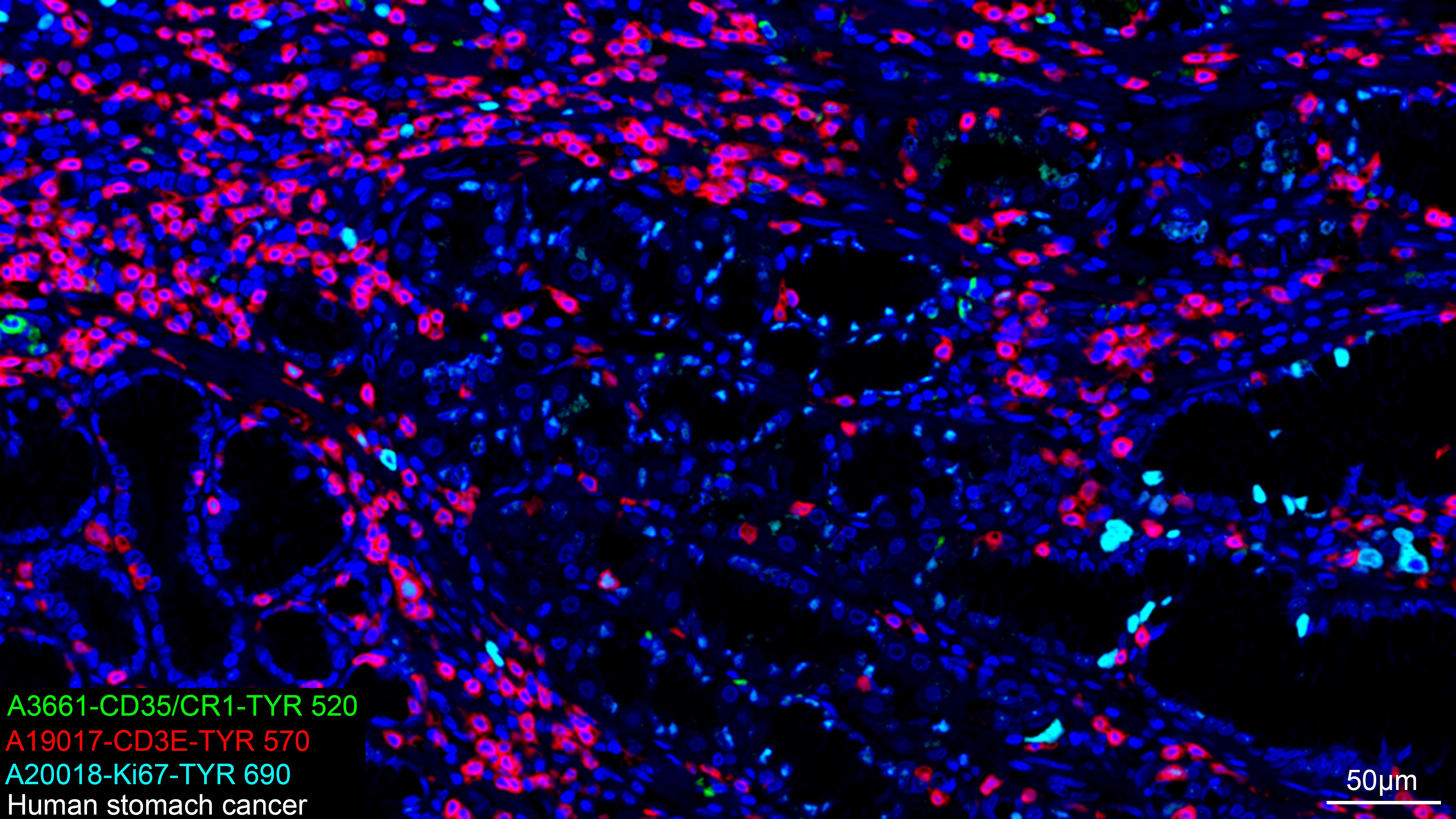
The multiplex IHC analysis on paraffin-embedded Human stomach cancer tissue using the following specific primary antibodies and tyramide signal amplification (TSA) reagents (RK05903) : CD35/CR1 Rabbit mAb (A3661, 1:100) with TSA-TYR-520 (Green), CD3E Rabbit mAb (CAB19017, 1:2000) with TSA-TYR-570 (Red), and Ki67 Rabbit mAb (A20018, 1:500) with TSA-TYR-690 (cyan). DAPI (Blue) was used for nuclear staining. Prior to multiplex IHC staining, high-pressure antigen retrieval was performed using 0.01M citrate buffer at pH 6.0. The analysis was completed using a 20x objective lens.
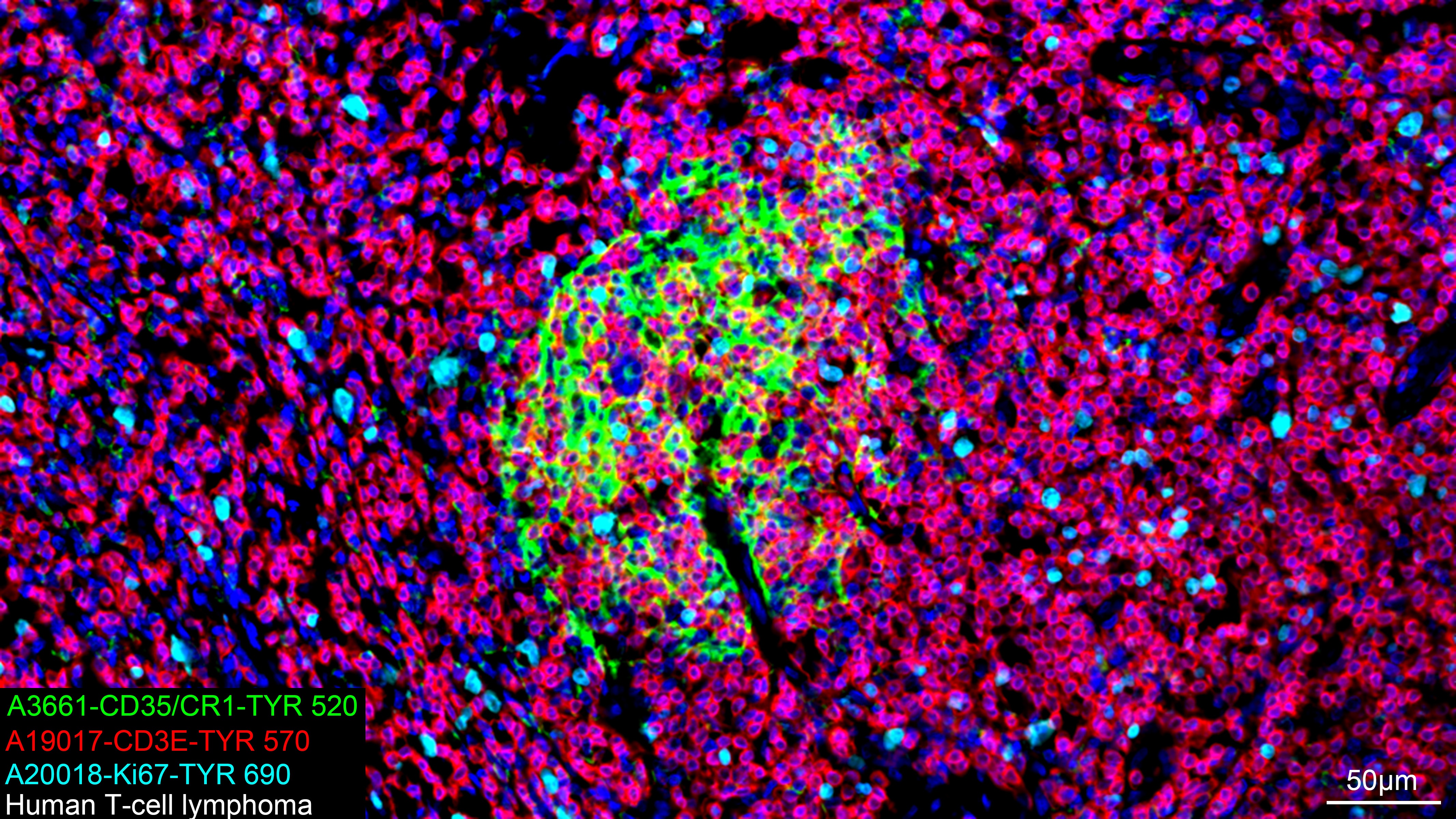
The multiplex IHC analysis on paraffin-embedded Human T-cell lymphoma tissue using the following specific primary antibodies and tyramide signal amplification (TSA) reagents (RK05903) : CD35/CR1 Rabbit mAb (A3661, 1:100) with TSA-TYR-520 (Green), CD3E Rabbit mAb (CAB19017, 1:2000) with TSA-TYR-570 (Red), and Ki67 Rabbit mAb (A20018, 1:500) with TSA-TYR-690 (cyan). DAPI (Blue) was used for nuclear staining. Prior to multiplex IHC staining, high-pressure antigen retrieval was performed using 0.01M citrate buffer at pH 6.0. The analysis was completed using a 20x objective lens.
The multiplex IHC analysis on paraffin-embedded Human T-cell lymphoma tissue using the following specific primary antibodies and tyramide signal amplification (TSA) reagents (RK05903) : CD35/CR1 Rabbit mAb (A3661, 1:100) with TSA-TYR-520 (Green), CD3E Rabbit mAb (CAB19017, 1:2000) with TSA-TYR-570 (Red), and Ki67 Rabbit mAb (A20018, 1:500) with TSA-TYR-690 (cyan). DAPI (Blue) was used for nuclear staining. Prior to multiplex IHC staining, high-pressure antigen retrieval was performed using 0.01M citrate buffer at pH 6.0. The analysis was completed using a 20x objective lens.


 (AGIM0524)")


