The CD34 Monoclonal Antibody (CAB19015) is a high-quality antibody developed for reliable detection and analysis of target proteins. This antibody, produced in rabbits, is highly specific to human CD34 and has been validated for use in various research applications, including immunohistochemistry and flow cytometry.CD34 is widely used in the field of hematology and stem cell research for its ability to identify and isolate stem cells from peripheral blood, bone marrow, and other sources. The CD34 Rabbit Monoclonal Antibody enables researchers to accurately detect CD34 expression in different cell types and tissues, providing valuable insights into stem cell biology, hematopoiesis, and regenerative medicine.
This antibody is validated for use in WB, IHC-P, ELISA, mIHC applications and has demonstrated reactivity against Human, Mouse, Rat samples.
Product Name:
CD34 Monoclonal Antibody
SKU:
CAB19015
Size:
20μL, 100μL
Reactivity:
Human, Mouse, Rat
Clone Number:
ARC0219
Conjugate:
Unconjugated
Immunogen:
Synthetic peptide. This information is considered to be commercially sensitive.
Recommended starting concentration is 1 μg/mL. Please optimize the concentration based on your specific assay requirements.
Synonyms:
CD34, CD34 molecule, GIG3, MORT1
Positive Sample:
bEnd.3, NIH/3T3, TF-1, TF-1 treated with PNGase F, NIH/3T3 treated with PNGase F
Cellular Localization:
Membrane, Single-Pass Type I Membrane Protein.
Calculated MW:
41kDa
Observed MW:
80-120kDa
The protein encoded by this gene may play a role in the attachment of stem cells to the bone marrow extracellular matrix or to stromal cells. This single-pass membrane protein is highly glycosylated and phosphorylated by protein kinase C. Two transcript variants encoding different isoforms have been found for this gene.
Purification Method
Affinity purification
Gene ID
947
RRID
AB_2862507
Buffer Information
Store at -20℃. Avoid freeze / thaw cycles. Buffer: PBS containing 50% glycerol and 0.05% BSA, preserved with proclin300 or sodium azide, pH 7.3.
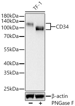
Western blot analysis of lysates from TF-1 cells using CD34 Rabbit mAb (CAB19015) at 1:1000 dilution incubated overnight at 4℃. TF-1 cells were treated with PNGase F (6 U/μL) at 37°C for 1.5 hours. Secondary antibody: HRP-conjugated Goat anti-Rabbit IgG (H+L) (CABS014) at 1:10000 dilution. Lysates/proteins: 25 μg per lane. Blocking buffer: 3% nonfat dry milk in TBST. Detection: ECL Basic Kit (AbGn00020). Exposure time: 90 s.
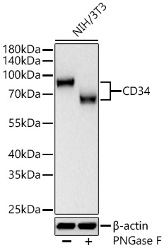
Western blot analysis of lysates from NIH/3T3 cells using CD34 Rabbit mAb (CAB19015) at 1:1000 dilution incubated overnight at 4℃. NIH/3T3 cells were treated with PNGase F (6 U/μL) at 37°C for 1.5 hours. Secondary antibody: HRP-conjugated Goat anti-Rabbit IgG (H+L) (CABS014) at 1:10000 dilution. Lysates/proteins: 25 μg per lane. Blocking buffer: 3% nonfat dry milk in TBST. Detection: ECL Basic Kit (AbGn00020). Exposure time: 90 s.
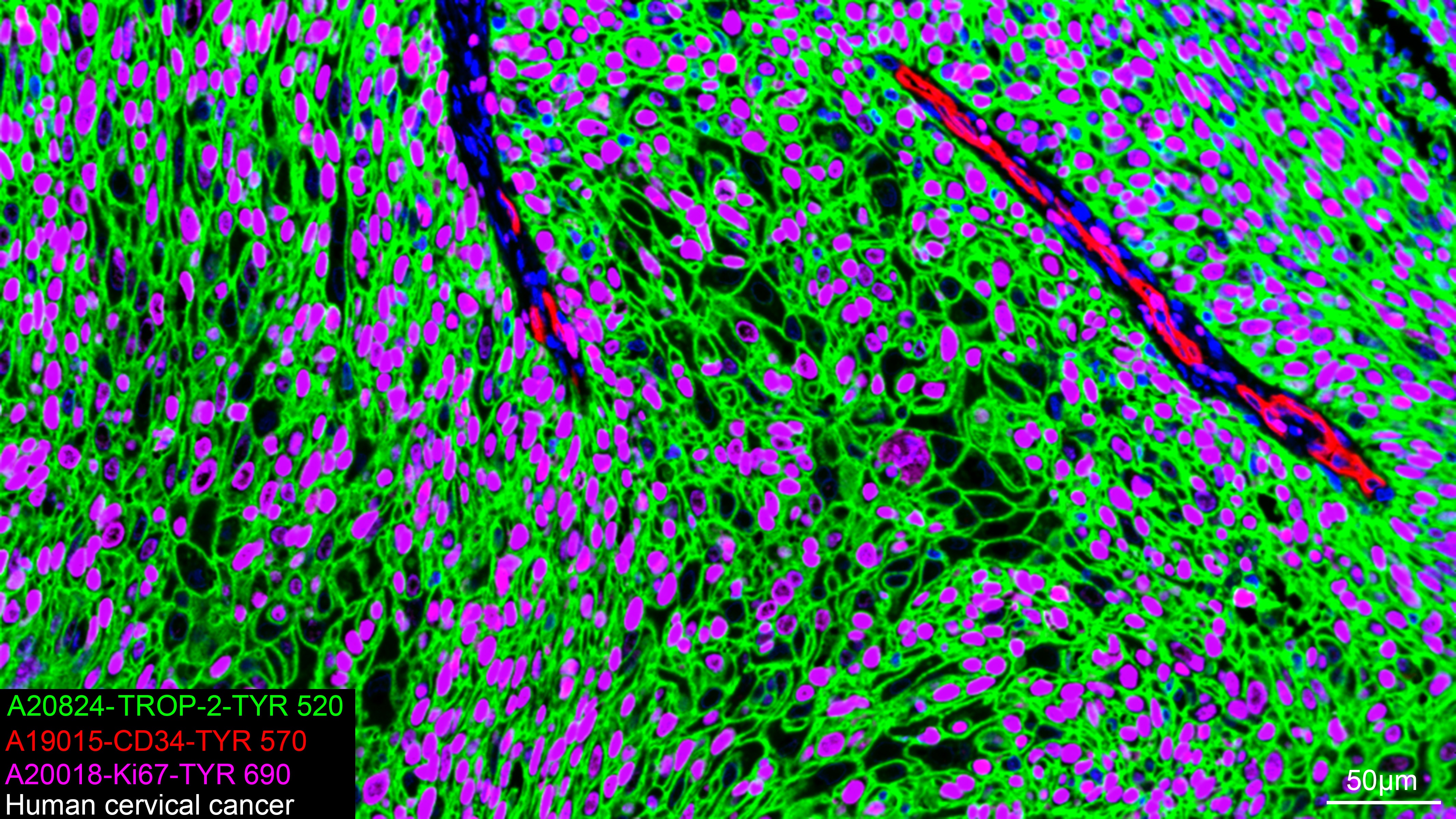
The multiplex IHC analysis on paraffin-embedded Human cervical cancer tissue using the following specific primary antibodies and tyramide signal amplification (TSA) reagents (RK05903) : TROP-2 Rabbit mAb (CAB20824, 1:8000) with TSA-TYR-520 (Green), CD34 Rabbit mAb (CAB19015, 1:100) with TSA-TYR-570 (Red), and Ki67 Rabbit mAb (CAB20018, 1:500) with TSA-TYR-690 (Magenta). DAPI (Blue) was used for nuclear staining. Prior to multiplex IHC staining, high-pressure antigen retrieval was performed using 0.01M citrate buffer at pH 6.0. The analysis was completed using a 20x objective lens.
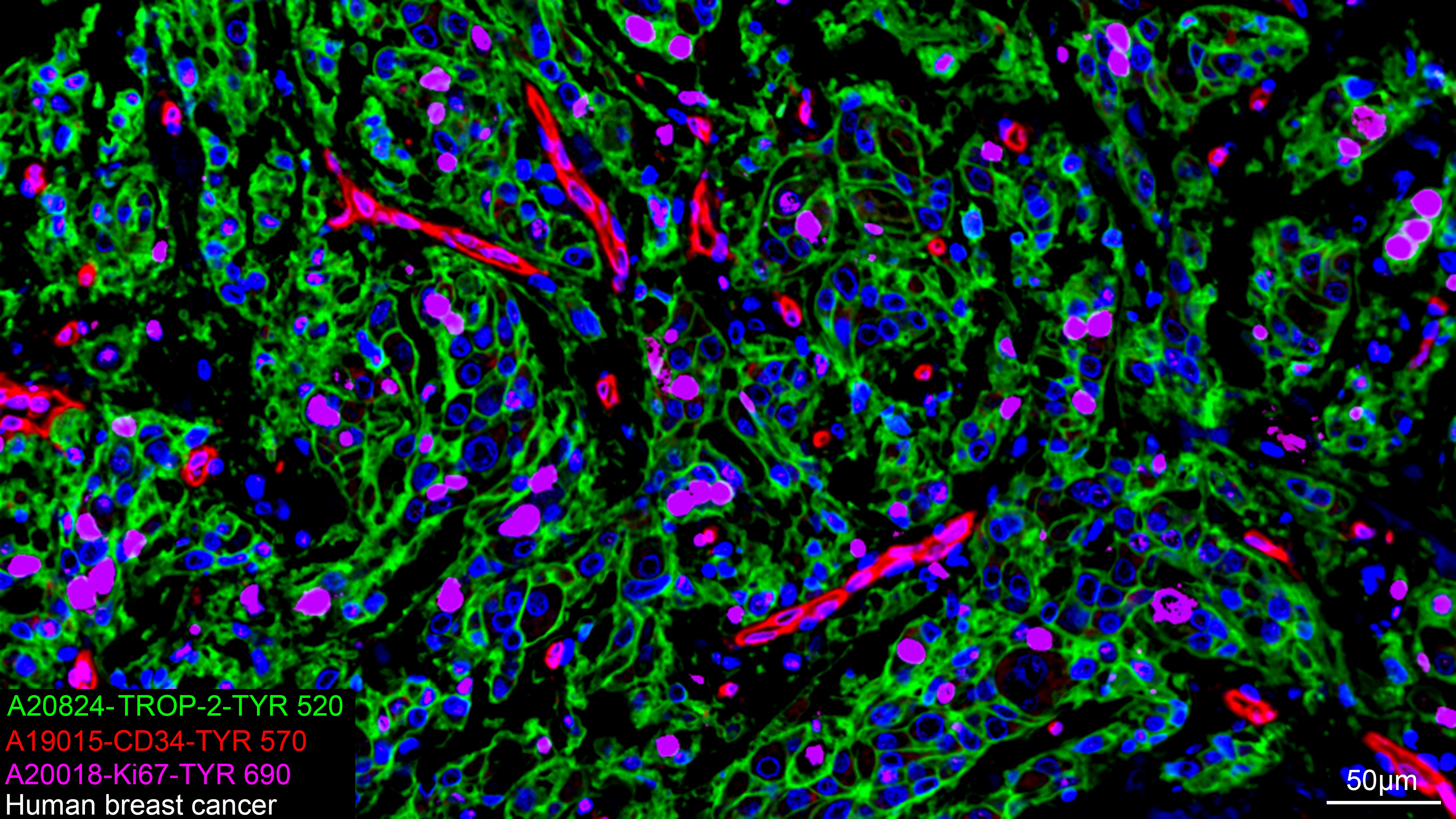
The multiplex IHC analysis on paraffin-embedded Human breast cancer tissue using the following specific primary antibodies and tyramide signal amplification (TSA) reagents (RK05903) : TROP-2 Rabbit mAb (CAB20824, 1:8000) with TSA-TYR-520 (Green), CD34 Rabbit mAb (CAB19015, 1:100) with TSA-TYR-570 (Red), and Ki67 Rabbit mAb (CAB20018, 1:500) with TSA-TYR-690 (Magenta). DAPI (Blue) was used for nuclear staining. Prior to multiplex IHC staining, high-pressure antigen retrieval was performed using 0.01M citrate buffer at pH 6.0. The analysis was completed using a 20x objective lens.