The CD7 Monoclonal Antibody (CAB9560) is a high-quality antibody developed for reliable detection and analysis of target proteins. This antibody, raised in rabbits, exhibits high specificity and sensitivity for detecting CD7 in human samples and is validated for use in various applications such as immunofluorescence and flow cytometry.CD7 plays a crucial role in T cell and NK cell development, activation, and function, making it a key target for research in immunology and cancer biology. By targeting CD7, researchers can better understand the mechanisms behind T cell and NK cell-mediated immune responses, ultimately leading to insights into immune-related diseases, including leukemia and lymphoma.
This antibody is validated for use in WB, IHC-P, ELISA applications and has demonstrated reactivity against Human samples.
Product Name:
CD7 Monoclonal Antibody
SKU:
CAB9560
Size:
20μL, 100μL
Reactivity:
Human
Clone Number:
ARC1634
Conjugate:
Unconjugated
Immunogen:
Synthetic peptide. This information is considered to be commercially sensitive.
Recommended starting concentration is 1 μg/mL. Please optimize the concentration based on your specific assay requirements.
Synonyms:
GP40, TP41, Tp40, LEU-9, CD7
Positive Sample:
Jurkat cells
Cellular Localization:
Membrane, Single-Pass Type I Membrane Protein.
Calculated MW:
25kDa
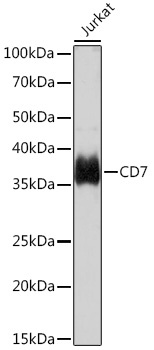
Observed MW:
37kDa
This gene encodes a transmembrane protein which is a member of the immunoglobulin superfamily. This protein is found on thymocytes and mature T cells. It plays an essential role in T-cell interactions and also in T-cell/B-cell interaction during early lymphoid development.
Purification Method
Affinity purification
Gene ID
924
RRID
AB_2863723
Buffer Information
Store at -20℃. Avoid freeze / thaw cycles. Buffer: PBS containing 50% glycerol and 0.05% BSA, preserved with proclin300 or sodium azide, pH 7.3.
Western blot analysis of lysates from Jurkat cells, using CD7 Rabbit mAb (CAB9560) at 1:1000 dilution. Secondary antibody: HRP-conjugated Goat anti-Rabbit IgG (H+L) (CABS014) at 1:10000 dilution. Lysates/proteins: 25μg per lane. Blocking buffer: 3% nonfat dry milk in TBST. Detection: ECL Basic Kit (AbGn00020). Exposure time: 30s.
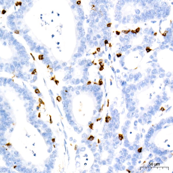
Immunohistochemistry analysis of paraffin-embedded Human colon carcinoma using CD7 Rabbit mAb (CAB9560) at dilution of 1:10000 (40x lens). High pressure antigen retrieval performed with 0.01M Tris/EDTA Buffer (pH 9.0) prior to IHC staining.