The CD79a Monoclonal Antibody (CAB19024) is a high-quality antibody developed for reliable detection and analysis of target proteins. This monoclonal antibody, produced in rabbits, exhibits high specificity and sensitivity for human samples, making it an ideal choice for Western blotting and immunohistochemistry applications.CD79A is a key regulator of B cell signaling and differentiation, playing a crucial role in antibody production and immune response. Dysregulation of CD79A expression or function is associated with various B cell-related disorders, including lymphomas and autoimmune diseases.
This antibody is validated for use in WB, IHC-P, ELISA applications and has demonstrated reactivity against Human samples.
Product Name:
CD79a Monoclonal Antibody
SKU:
CAB19024
Size:
20μL, 100μL
Reactivity:
Human
Clone Number:
ARC0482
Conjugate:
Unconjugated
Immunogen:
Synthetic peptide. This information is considered to be commercially sensitive.
Recommended starting concentration is 1 μg/mL. Please optimize the concentration based on your specific assay requirements.
Synonyms:
IGA, MB1, MB-1, IGAlpha, CD79a
Positive Sample:
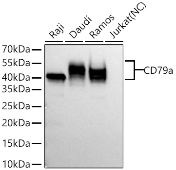
Raji, Daudi, Ramos
Cellular Localization:
Cell Membrane, Single-Pass Type I Membrane Protein.
Calculated MW:
25kDa
Observed MW:
40-55kDa
The B lymphocyte antigen receptor is a multimeric complex that includes the antigen-specific component, surface immunoglobulin (Ig). Surface Ig non-covalently associates with two other proteins, Ig-alpha and Ig-beta, which are necessary for expression and function of the B-cell antigen receptor. This gene encodes the Ig-alpha protein of the B-cell antigen component. Alternatively spliced transcript variants encoding different isoforms have been described.
Purification Method
Affinity purification
Gene ID
973
RRID
AB_2862516
Buffer Information
Store at -20℃. Avoid freeze / thaw cycles. Buffer: PBS containing 50% glycerol and 0.05% BSA, preserved with proclin300 or sodium azide, pH 7.3.
Western blot analysis of various lysates using CD79a Rabbit mAb (CAB19024) at 1:5000 dilution incubated at room temperature for 1.5 hours. Secondary antibody: HRP-conjugated Goat anti-Rabbit IgG (H+L) (CABS014) at 1:10000 dilution. Lysates/proteins: 25 μg per lane. Blocking buffer: 3% nonfat dry milk in TBST. Detection: ECL Basic Kit (AbGn00020). Negative control (NC): Jurkat Exposure time: 60s.
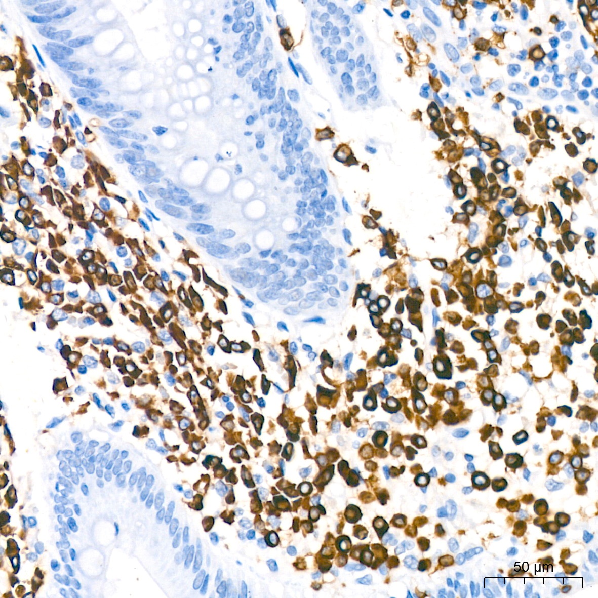
Immunohistochemistry analysis of paraffin-embedded Human appendix using CD79a Rabbit mAb (CAB19024) at dilution of 1:400 (40x lens). High pressure antigen retrieval performed with 0.01M Citrate buffer (pH 6.0) prior to IHC staining.
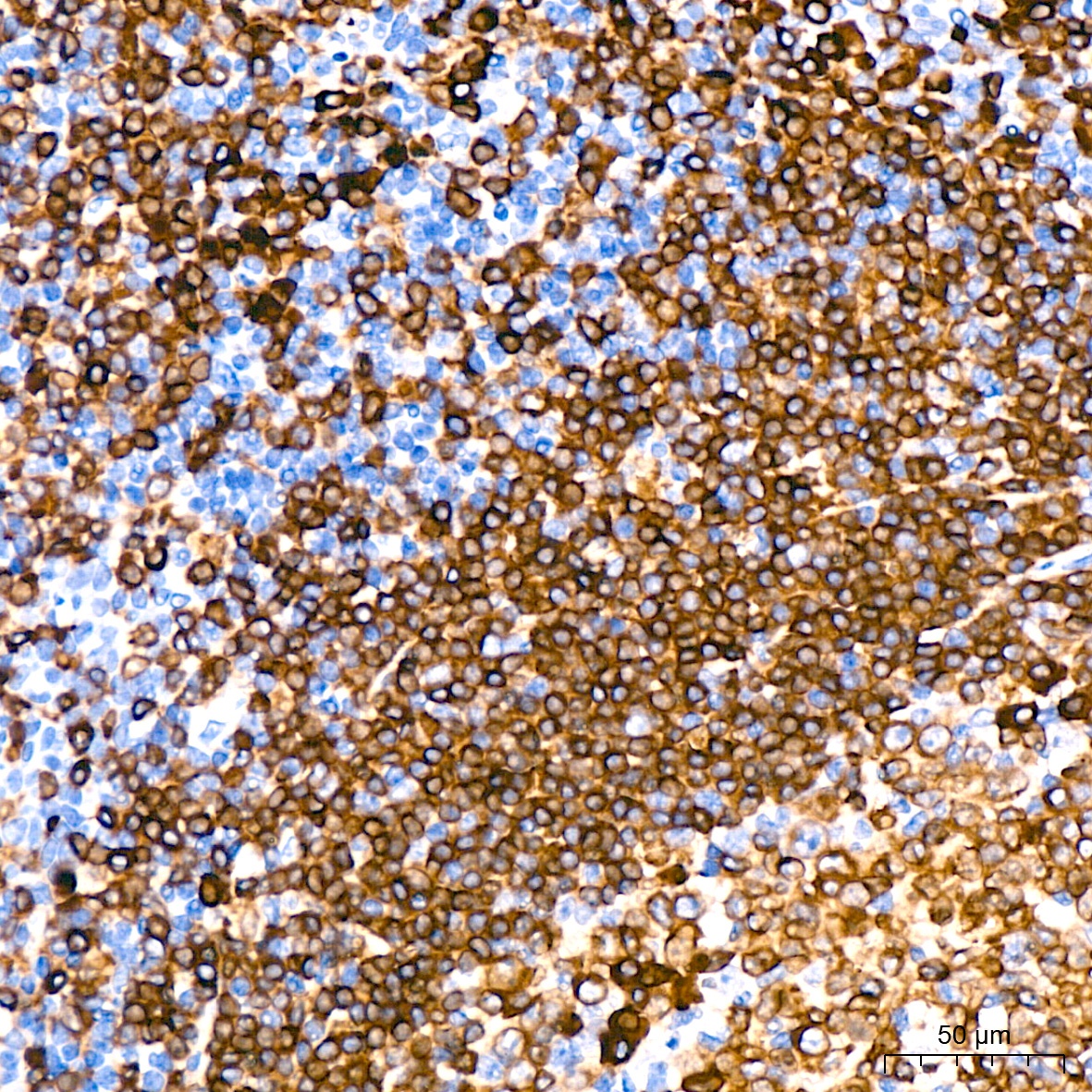
Immunohistochemistry analysis of paraffin-embedded Human tonsil using CD79a Rabbit mAb (CAB19024) at dilution of 1:400 (40x lens). High pressure antigen retrieval performed with 0.01M Citrate buffer (pH 6.0) prior to IHC staining.