The CDH5 Antibody (CAB12416) is a high-quality antibody developed for reliable detection and analysis of target proteins. This cell adhesion molecule plays a critical role in vascular development and maintenance, making it a key player in angiogenesis and endothelial cell function. Raised in rabbits, this antibody is highly reactive with human samples and has been validated for use in Western blot applications. It specifically binds to the CDH5 protein, enabling precise detection and analysis in various cell types and tissues. Research involving CDH5 is essential for understanding the mechanisms underlying vascular development, as well as for studying diseases such as cancer, cardiovascular disorders, and inflammatory conditions.
This antibody is validated for use in WB, IHC-P, ELISA applications and has demonstrated reactivity against Human, Mouse samples.
Product Name:
CDH5 Antibody
SKU:
CAB12416
Size:
20μL, 100μL
Reactivity:
Human, Mouse
Conjugate:
Unconjugated
Immunogen:
Recombinant protein (or fragment).This information is considered to be commercially sensitive.
Recommended starting concentration is 1 μg/mL. Please optimize the concentration based on your specific assay requirements.
Synonyms:
7B4, CD144, VE Cadherin
Positive Sample:
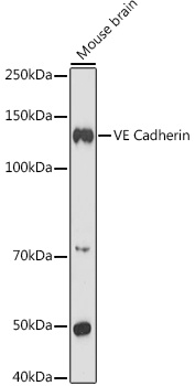
Mouse brain
Cellular Localization:
Cell Junction, Cell Membrane, Single-Pass Type I Membrane Protein.
Calculated MW:
88kDa
Observed MW:
140kDa
This gene encodes a classical cadherin of the cadherin superfamily. The encoded preproprotein is proteolytically processed to generate the mature glycoprotein. This calcium-dependent cell-cell adhesion molecule is comprised of five extracellular cadherin repeats, a transmembrane region and a highly conserved cytoplasmic tail. Functioning as a classical cadherin by imparting to cells the ability to adhere in a homophilic manner, this protein plays a role in endothelial adherens junction assembly and maintenance. This gene is located in a gene cluster in a region on the long arm of chromosome 16 that is involved in loss of heterozygosity events in breast and prostate cancer.
Purification Method
Affinity purification
Gene ID
1003
RRID
AB_2759258
Buffer Information
Store at -20℃. Avoid freeze / thaw cycles. Buffer: PBS containing 50% glycerol, preserved with proclin300 or sodium azide, pH 7.3.
Western blot analysis of lysates from Mouse brain, using VE Cadherin Rabbit pAb (CAB12416) at 1:1000 dilution. Secondary antibody: HRP-conjugated Goat anti-Rabbit IgG (H+L) (CABS014) at 1:10000 dilution. Lysates/proteins: 25μg per lane. Blocking buffer: 3% nonfat dry milk in TBST. Detection: ECL Enhanced Kit (AbGn00021). Exposure time: 180s.
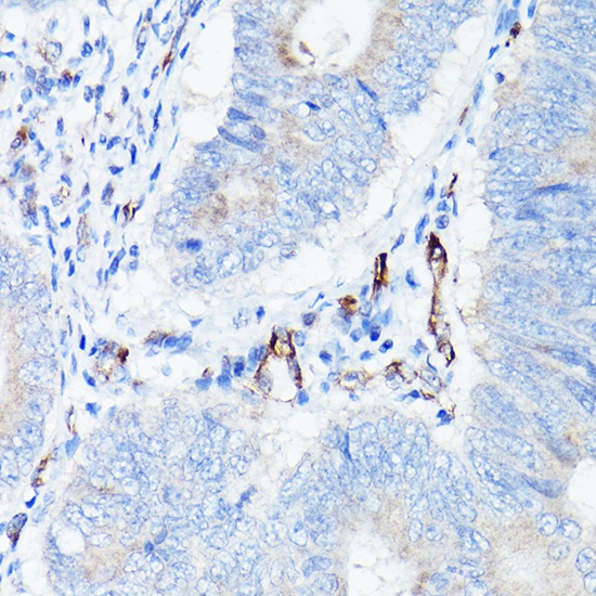
Immunohistochemistry analysis of paraffin-embedded Human colon carcinoma using VE Cadherin Rabbit pAb (CAB12416) at dilution of 1:100 (40x lens). High pressure antigen retrieval performed with 0.01M Citrate buffer (pH 6.0) prior to IHC staining.