The CDH9 Antibody (CAB14716) is a high-quality antibody developed for reliable detection and analysis of target proteins. This antibody, produced in rabbits, has high reactivity with human samples and is validated for use in various applications including Western blot, immunofluorescence, and immunohistochemistry.CDH9, also known as cadherin-9, plays a crucial role in neural cell adhesion and communication, making it a key player in the formation and maintenance of neural circuits. Its expression in the brain and spinal cord suggests its involvement in neurological disorders and developmental abnormalities.
This antibody is validated for use in WB, ELISA applications and has demonstrated reactivity against Human samples.
Product Name:
CDH9 Antibody
SKU:
CAB14716
Size:
20μL, 100μL
Reactivity:
Human
Conjugate:
Unconjugated
Immunogen:
Recombinant protein (or fragment).This information is considered to be commercially sensitive.
Recommended starting concentration is 1 μg/mL. Please optimize the concentration based on your specific assay requirements.
Synonyms:
CDH9
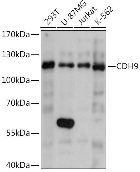
Positive Sample:
293T, U-87MG, Jurkat, K-562
Cellular Localization:
Cell Membrane, Single-Pass Type I Membrane Protein.
Calculated MW:
89kDa
Observed MW:
120kDa
This gene encodes a type II classical cadherin from the cadherin superfamily, integral membrane proteins that mediate calcium-dependent cell-cell adhesion. Mature cadherin proteins are composed of a large N-terminal extracellular domain, a single membrane-spanning domain, and a small, highly conserved C-terminal cytoplasmic domain. The extracellular domain consists of 5 subdomains, each containing a cadherin motif, and appears to determine the specificity of the protein's homophilic cell adhesion activity. Type II (atypical) cadherins are defined based on their lack of a HAV cell adhesion recognition sequence specific to type I cadherins.
Purification Method
Affinity purification
Gene ID
1007
RRID
AB_2761592
Buffer Information
Store at -20℃. Avoid freeze / thaw cycles. Buffer: PBS with 0.01% thimerosal,50% glycerol,pH7.3.
Western blot analysis of various lysates using CDH9 Rabbit pAb (CAB14716) at 1:1000 dilution. Secondary antibody: HRP-conjugated Goat anti-Rabbit IgG (H+L) (CABS014) at 1:10000 dilution. Lysates/proteins: 25μg per lane. Blocking buffer: 3% nonfat dry milk in TBST. Detection: ECL Basic Kit (AbGn00020). Exposure time: 10s.