The CDX2 Monoclonal Antibody (CAB19030) is a high-quality antibody developed for reliable detection and analysis of target proteins. This antibody, derived from rabbit monoclonal cells, exhibits high specificity and sensitivity towards human CDX2 protein, making it ideal for various research applications such as immunohistochemistry and flow cytometry.CDX2 is known for its role in maintaining the integrity of the intestinal epithelium and regulating the expression of genes involved in gut development.
This antibody is validated for use in WB, IHC-P, IP, ELISA, IF-P applications and has demonstrated reactivity against Human, Mouse, Rat samples.
Product Name:
CDX2 Monoclonal Antibody
SKU:
CAB19030
Size:
20μL, 100μL
Reactivity:
Human, Mouse, Rat
Clone Number:
ARC0450
Conjugate:
Unconjugated
Immunogen:
Synthetic peptide. This information is considered to be commercially sensitive.
0.5μg-4μg antibody for 200μg-600μg extracts of whole cells
IF-P
1:100 - 1:1000
IHC-P
1:3000 - 1:12000
ELISA
Recommended starting concentration is 1 μg/mL. Please optimize the concentration based on your specific assay requirements.
Synonyms:
CDX3, CDX-3, CDX2/AS, X2
Positive Sample:
SW480, 293T
Cellular Localization:
Nucleus.
Calculated MW:
34kDa
Observed MW:
38kDa
This gene is a member of the caudal-related homeobox transcription factor gene family. The encoded protein is a major regulator of intestine-specific genes involved in cell growth an differentiation. This protein also plays a role in early embryonic development of the intestinal tract. Aberrant expression of this gene is associated with intestinal inflammation and tumorigenesis.
Purification Method
Affinity purification
Gene ID
1045
RRID
AB_2862522
Buffer Information
Store at -20℃. Avoid freeze / thaw cycles. Buffer: PBS containing 50% glycerol and 0.05% BSA, preserved with proclin300 or sodium azide, pH 7.3.
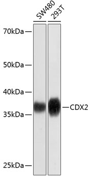
Western blot analysis of various lysates using [KD Validated] CDX2 Rabbit mAb (CAB19030) at 1:1000 dilution. Secondary antibody: HRP-conjugated Goat anti-Rabbit IgG (H+L) (CABS014) at 1:10000 dilution. Lysates/proteins: 25μg per lane. Blocking buffer: 3% nonfat dry milk in TBST. Detection: ECL Basic Kit (AbGn00020). Exposure time: 3min.
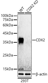
Western blot analysis of lysates from wild type(WT) and CDX2 knockdown (KD) 293T cells, using [KD Validated] CDX2 Rabbit mAb (CAB19030) at 1:500 dilution. Secondary antibody: HRP-conjugated Goat anti-Rabbit IgG (H+L) (CABS014) at 1:10000 dilution. Lysates/proteins: 25μg per lane. Blocking buffer: 3% nonfat dry milk in TBST. Detection: ECL Basic Kit (AbGn00020). Exposure time: 30s.
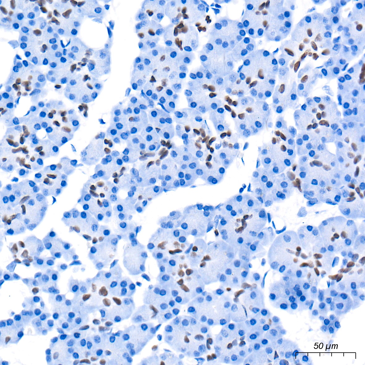
Immunohistochemistry analysis of paraffin-embedded Human pancreas tissue using [KD Validated] CDX2 Rabbit mAb (CAB19030) at a dilution of 1:4000 (40x lens). High pressure antigen retrieval performed with 0.01M Tris-EDTA Buffer (pH 9.0) prior to IHC staining.
Immunohistochemistry analysis of paraffin-embedded Mouse colon tissue using [KD Validated] CDX2 Rabbit mAb (CAB19030) at a dilution of 1:4000 (40x lens). High pressure antigen retrieval performed with 0.01M Tris-EDTA Buffer (pH 9.0) prior to IHC staining.
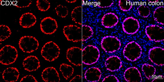
Confocal imaging of Human colon using [KD Validated] CDX2 Rabbit mAb (CAB19030,dilution 1:100)(Red). DAPI was used for nuclear staining (blue). Objective: 40x.
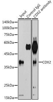
Immunoprecipitation analysis of 600 μg extracts of 293T cells using 3 μg [KD Validated] CDX2 Rabbit mAb (CAB19030). Western blot was performed from the immunoprecipitate using CDX2 antibody (CAB19030) at a dilution of 1:500.