The Chk2 Antibody (CAB0466) is a high-quality antibody developed for reliable detection and analysis of target proteins. In response to DNA damage and replication blocks, cell cycle progression is halted through the control of critical cell cycle regulators. The protein encoded by this gene is a cell cycle checkpoint regulator and putative tumor suppressor. It contains a forkhead-associated protein interaction domain essential for activation in response to DNA damage and is rapidly phosphorylated in response to replication blocks and DNA damage. When activated, the encoded protein is known to inhibit CDC25C phosphatase, preventing entry into mitosis, and has been shown to stabilize the tumor suppressor protein p53, leading to cell cycle arrest in G1. In addition, this protein interacts with and phosphorylates BRCA1, allowing BRCA1 to restore survival after DNA damage. Mutations in this gene have been linked with Li-Fraumeni syndrome, a highly penetrant familial cancer phenotype usually associated with inherited mutations in TP53. Also, mutations in this gene are thought to confer a predisposition to sarcomas, breast cancer, and brain tumors. This nuclear protein is a member of the CDS1 subfamily of serine/threonine protein kinases. Several transcript variants encoding different isoforms have been found for this gene. RRID AB_2757206 Gene ID 11200 Swiss Prot Synonym CDS1; CHK2; LFS2; RAD53; hCds1; HuCds1; PP1425; Chk2
This antibody is validated for use in WB, IF/ICC, IP, ELISA applications and has demonstrated reactivity against Human samples.
Product Name:
Chk2 Antibody
SKU:
CAB0466
Size:
100μL, 20μL
Reactivity:
Human
Clone Number:
-
Conjugate:
Unconjugated
Immunogen:
Recombinant protein (or fragment).This information is considered to be commercially sensitive.
Tested Applications:
WBIF/ICCIPELISA
Recommended Dilution:
WB
1:500 - 1:1000
IF
/
ICC
1:50 - 1:200
IP
0.5μg-4μg antibody for 200μg-400μg extracts of whole cells
ELISA
Recommended starting concentration is 1 μg/mL. Please optimize the concentration based on your specific assay requirements.
In response to DNA damage and replication blocks, cell cycle progression is halted through the control of critical cell cycle regulators. The protein encoded by this gene is a cell cycle checkpoint regulator and putative tumor suppressor. It contains a forkhead-associated protein interaction domain essential for activation in response to DNA damage and is rapidly phosphorylated in response to replication blocks and DNA damage. When activated, the encoded protein is known to inhibit CDC25C phosphatase, preventing entry into mitosis, and has been shown to stabilize the tumor suppressor protein p53, leading to cell cycle arrest in G1. In addition, this protein interacts with and phosphorylates BRCA1, allowing BRCA1 to restore survival after DNA damage. Mutations in this gene have been linked with Li-Fraumeni syndrome, a highly penetrant familial cancer phenotype usually associated with inherited mutations in TP53. Also, mutations in this gene are thought to confer a predisposition to sarcomas, breast cancer, and brain tumors. This nuclear protein is a member of the CDS1 subfamily of serine/threonine protein kinases. Several transcript variants encoding different isoforms have been found for this gene. RRID AB_2757206 Gene ID 11200 Swiss Prot Synonym CDS1; CHK2; LFS2; RAD53; hCds1; HuCds1; PP1425; Chk2
Purification Method:
Affinity purification
Gene ID:
11200
RRID:
AB_2757206
Buffer Information:
Store at -20℃. Avoid freeze / thaw cycles. Buffer: PBS containing 50% glycerol, preserved with proclin300 or sodium azide, pH 7.3.
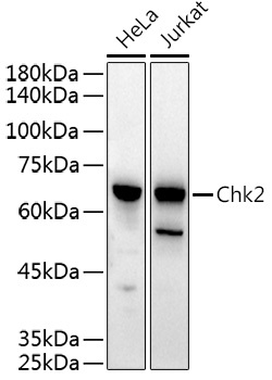
Western blot analysis of various lysates using Chk2 Rabbit pAb (CAB0466) at 1:500 dilution. Secondary antibody: HRP-conjugated Goat anti-Rabbit IgG (H+L) (AS014) at 1:10000 dilution. Lysates/proteins: 25μg per lane. Blocking buffer: 3% nonfat dry milk in TBST. Detection: ECL Basic Kit (AbGn00020). Exposure time: 30s.
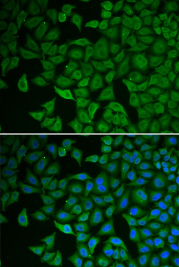
Immunofluorescence analysis of HeLa cells using Chk2 Rabbit pAb (CAB0466). Secondary antibody: Cy3-conjugated Goat anti-Rabbit IgG (H+L) (AS007) at 1:500 dilution. Blue: DAPI for nuclear staining.
Immunoprecipitation analysis of 200 μg extracts of MCF-7 cells using 1 μg Chk2 antibody (CAB0466). Western blot was performed from the immunoprecipitate using Chk2 antibody (CAB0466) at a dilution of 1:1000.