The CHRM1 Antibody (CAB16819) is a high-quality antibody developed for reliable detection and analysis of target proteins. This antibody, derived from rabbit serum, is highly specific for human samples and is validated for use in Western blot applications. By binding to the CHRM1 protein, the antibody enables accurate detection and analysis in a variety of cell types, making it ideal for studies in neurology, pharmacology, and drug development.CHRM1 is an important receptor in the central and peripheral nervous systems, where it plays a crucial role in mediating the effects of acetylcholine.
This antibody is validated for use in WB, IF/ICC, ELISA applications and has demonstrated reactivity against Human, Mouse, Rat samples.
Product Name:
CHRM1 Antibody
SKU:
CAB16819
Size:
20μL, 100μL
Reactivity:
Human, Mouse, Rat
Immunogen:
Synthetic peptide. This information is considered to be commercially sensitive.
The muscarinic cholinergic receptors belong to a larger family of G protein-coupled receptors. The functional diversity of these receptors is defined by the binding of acetylcholine and includes cellular responses such as adenylate cyclase inhibition, phosphoinositide degeneration, and potassium channel mediation. Muscarinic receptors influence many effects of acetylcholine in the central and peripheral nervous system. The muscarinic cholinergic receptor 1 is involved in mediation of vagally-induced bronchoconstriction and in the acid secretion of the gastrointestinal tract. The gene encoding this receptor is localized to 11q13.
Purification Method
Affinity purification
Gene ID
1128
RRID
AB_2768907
Buffer Information
Store at -20℃. Avoid freeze / thaw cycles. Buffer: PBS with 0.01% thimerosal,50% glycerol,pH7.3.
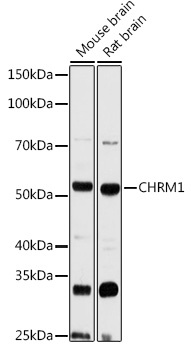
Western blot analysis of various lysates using CHAbGn1 Rabbit pAb (CAB16819) at 1:3000 dilution. Secondary antibody: HRP-conjugated Goat anti-Rabbit IgG (H+L) (CABS014) at 1:10000 dilution. Lysates/proteins: 25μg per lane. Blocking buffer: 3% nonfat dry milk in TBST. Detection: ECL Basic Kit (AbGn00020). Exposure time: 90s.
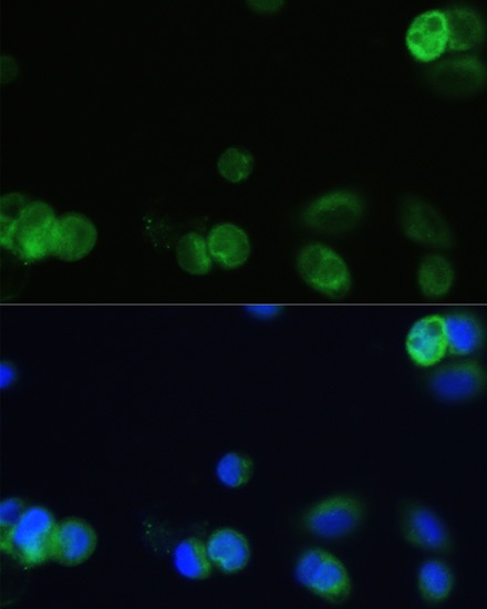
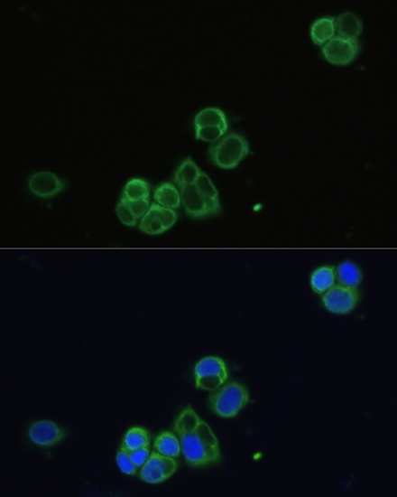
Immunofluorescence analysis of OVCAR-3 cells using CHAbGn1 Rabbit pAb (CAB16819) at dilution of 1:100. Secondary antibody: Cy3-conjugated Goat anti-Rabbit IgG (H+L) (CABS007) at 1:500 dilution. Blue: DAPI for nuclear staining.
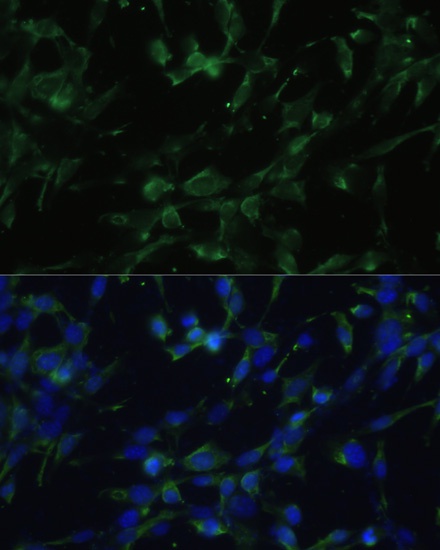
Immunofluorescence analysis of NIH-3T3 cells using CHAbGn1 Rabbit pAb (CAB16819) at dilution of 1:100. Secondary antibody: Cy3-conjugated Goat anti-Rabbit IgG (H+L) (CABS007) at 1:500 dilution. Blue: DAPI for nuclear staining.
Immunofluorescence analysis of OVCAR-3 cells using CHAbGn1 Rabbit pAb (CAB16819) at dilution of 1:100. Secondary antibody: Cy3-conjugated Goat anti-Rabbit IgG (H+L) (CABS007) at 1:500 dilution. Blue: DAPI for nuclear staining.