The cIAP1 Monoclonal Antibody (CAB19688) is a high-quality antibody developed for reliable detection and analysis of target proteins. This antibody is raised in rabbits and is highly specific for CIAP1 in human samples, making it an ideal choice for Western blot applications. By targeting CIAP1, researchers can detect and analyze this important protein in various cell types, allowing for further insights into its role in immunology and cancer research.CIAP1, also known as cellular inhibitor of apoptosis protein 1, is a key player in regulating cell survival and apoptosis, making it a promising target for studying diseases such as cancer and inflammatory disorders.
This antibody is validated for use in WB, IHC-P, ELISA applications and has demonstrated reactivity against Human, Rat samples.
Product Name:
cIAP1 Monoclonal Antibody
SKU:
CAB19688
Size:
20μL, 100μL
Reactivity:
Human, Rat
Clone Number:
ARC0168
Conjugate:
Unconjugated
Immunogen:
Synthetic peptide. This information is considered to be commercially sensitive.
The protein encoded by this gene is a member of a family of proteins that inhibits apoptosis by binding to tumor necrosis factor receptor-associated factors TRAF1 and TRAF2, probably by interfering with activation of ICE-like proteases. This encoded protein inhibits apoptosis induced by serum deprivation and menadione, a potent inducer of free radicals. Alternatively spliced transcript variants encoding different isoforms have been found for this gene.
Purification Method
Affinity purification
Gene ID
329
RRID
AB_2862736
Buffer Information
Store at -20℃. Avoid freeze / thaw cycles. Buffer: PBS containing 50% glycerol and 0.05% BSA, preserved with proclin300 or sodium azide, pH 7.3.
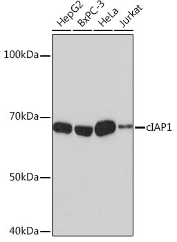
Western blot analysis of various lysates using [KO Validated] cIAP1 Rabbit mAb (CAB19688) at 1:1000 dilution. Secondary antibody: HRP-conjugated Goat anti-Rabbit IgG (H+L) (CABS014) at 1:10000 dilution. Lysates/proteins: 25μg per lane. Blocking buffer: 3% nonfat dry milk in TBST. Detection: ECL Basic Kit (AbGn00020). Exposure time: 3min.
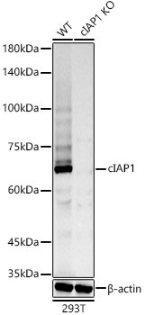
Western blot analysis of lysates from wild type(WT) and cIAP1 knockout (KO) 293T cells, using [KO Validated] cIAP1 Rabbit mAb (CAB19688) at 1:1000 dilution. Secondary antibody: HRP-conjugated Goat anti-Rabbit IgG (H+L) (CABS014) at 1:10000 dilution. Lysates/proteins: 25μg per lane. Blocking buffer: 3% nonfat dry milk in TBST. Detection: ECL Basic Kit (AbGn00020). Exposure time: 90s.