The CIDEB Antibody (CAB17140) is a high-quality antibody developed for reliable detection and analysis of target proteins. This antibody, generated in rabbits, exhibits high specificity and sensitivity for detecting CIDEB in human samples, making it ideal for use in Western blot applications. By targeting CIDEB, researchers can gain insights into its role in lipid homeostasis and inflammatory processes, offering potential applications in studies of metabolic disorders, liver diseases, and immune-related conditions.CIDEB, a key player in lipid droplet formation and lipid metabolism, is implicated in various physiological and pathological processes, including lipid accumulation, insulin resistance, and inflammation.
This antibody is validated for use in WB, ELISA applications and has demonstrated reactivity against Human samples.
Product Name:
CIDEB Antibody
SKU:
CAB17140
Size:
20μL, 100μL
Reactivity:
Human
Immunogen:
Recombinant protein (or fragment).This information is considered to be commercially sensitive.
Recommended starting concentration is 1 μg/mL. Please optimize the concentration based on your specific assay requirements.
Synonyms:
CIDEB, cell death activator CIDE-B, IN, LHR, MC56, MDU2, MDU3, MIC4, Pgp1, CDW44, CSPG8, HCELL, HUTCH-I, ECMR-III
Positive Sample:
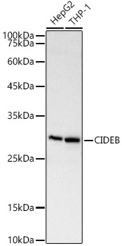
HepG2, THP-1
Cellular Localization:
Cytosol, Perinuclear Region Of Cytoplasm.
Calculated MW:
25kDa
Observed MW:
28kDa
Enables identical protein binding activity. Involved in activation of cysteine-type endopeptidase activity; positive regulation of cell death; and positive regulation of release of cytochrome c from mitochondria. Acts upstream of or within apoptotic process. Located in cytosol and perinuclear region of cytoplasm.
Purification Method
Affinity purification
Gene ID
27141
RRID
AB_2768923
Buffer Information
Store at -20℃. Avoid freeze / thaw cycles. Buffer: Buffer: PBS containing 50% glycerol, preserved with proclin300 or sodium azide, pH 7.3.
Western blot analysis of various lysates, using CIDEB Rabbit pAb (CAB17140) at 1:2000 dilution. Secondary antibody: HRP-conjugated Goat anti-Rabbit IgG (H+L) (CABS014) at 1:10000 dilution. Lysates/proteins: 25μg per lane. Blocking buffer: 3% nonfat dry milk in TBST. Detection: ECL Basic Kit (AbGn00020). Exposure time: 10s.