The CLN5 Antibody (CAB12886) is a high-quality antibody developed for reliable detection and analysis of target proteins. This antibody, produced in rabbits, exhibits high reactivity with human samples and has been validated for use in Western blot applications. By binding specifically to the CLN5 protein, this antibody enables the detection and analysis of CLN5 in various cell types, making it ideal for investigations in neurology and genetic disorders.
This antibody is validated for use in WB, IHC-P, ELISA applications and has demonstrated reactivity against Human, Mouse, Rat samples.
Product Name:
CLN5 Antibody
SKU:
CAB12886
Size:
20μL, 100μL
Reactivity:
Human, Mouse, Rat
Conjugate:
Unconjugated
Immunogen:
Recombinant protein (or fragment).This information is considered to be commercially sensitive.
Recommended starting concentration is 1 μg/mL. Please optimize the concentration based on your specific assay requirements.
Synonyms:
CLN5, NCL
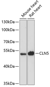
Positive Sample:
Mouse heart, Rat heart
Cellular Localization:
Lysosome.
Calculated MW:
41kDa
Observed MW:
41kDa
This gene is one of eight which have been associated with neuronal ceroid lipofuscinoses (NCL). Also referred to as Batten disease, NCL comprises a class of autosomal recessive, neurodegenerative disorders affecting children. The genes responsible likely encode proteins involved in the degradation of post-translationally modified proteins in lysosomes. The primary defect in NCL disorders is thought to be associated with lysosomal storage function.
Purification Method
Affinity purification
Gene ID
1203
RRID
AB_2759729
Buffer Information
Store at -20℃. Avoid freeze / thaw cycles. Buffer: PBS with 0.01% thimerosal,50% glycerol,pH7.3.
Western blot analysis of various lysates using CLN5 Rabbit pAb (CAB12886) at 1:3000 dilution. Secondary antibody: HRP-conjugated Goat anti-Rabbit IgG (H+L) (CABS014) at 1:10000 dilution. Lysates/proteins: 25μg per lane. Blocking buffer: 3% nonfat dry milk in TBST. Detection: ECL Basic Kit (AbGn00020). Exposure time: 90s.
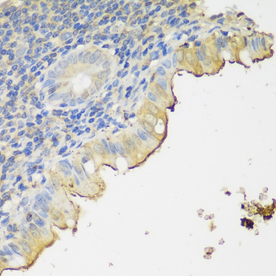
Immunohistochemistry analysis of paraffin-embedded Human appendix using CLN5 Rabbit pAb (CAB12886) at dilution of 1:150 (40x lens). Microwave antigen retrieval performed with 0.01M PBS Buffer (pH 7.2) prior to IHC staining.