The Mast Cell Chymase (CMA1) Monoclonal Antibody (CAB11480) is a high-quality antibody developed for reliable detection and analysis of target proteins. This antibody, produced in rabbits, shows high reactivity with human samples and is validated for use in applications such as immunohistochemistry and ELISA.Mast cell chymase is a key player in immune responses, tissue remodeling, and inflammation regulation. Its dysregulation has been linked to conditions like asthma, cardiovascular diseases, and cancer. By targeting mast cell chymase, researchers can gain valuable insights into disease mechanisms and potentially develop new therapeutic strategies.
This antibody is validated for use in WB, IHC-P, ELISA applications and has demonstrated reactivity against Human, Mouse, Rat samples.
Product Name:
Mast Cell Chymase (CMA1) Monoclonal Antibody
SKU:
CAB11480
Size:
20μL, 100μL
Reactivity:
Human, Mouse, Rat
Clone Number:
ARC0614
Conjugate:
Unconjugated
Immunogen:
Synthetic peptide. This information is considered to be commercially sensitive.
Recommended starting concentration is 1 μg/mL. Please optimize the concentration based on your specific assay requirements.
Synonyms:
CYH, MCT1, chymase, Mast Cell Chymase (CMA1)
Positive Sample:
A549, Mouse heart, Rat lung, Rat small intestine
Cellular Localization:
Cytoplasmic Granule, Secreted.
Calculated MW:
27kDa
Observed MW:
27kDa
This gene encodes a chymotryptic serine proteinase that belongs to the peptidase family S1. It is expressed in mast cells and is thought to function in the degradation of the extracellular matrix, the regulation of submucosal gland secretion, and the generation of vasoactive peptides. In the heart and blood vessels, this protein, rather than angiotensin converting enzyme, is largely responsible for converting angiotensin I to the vasoactive peptide angiotensin II. Alternative splicing results in multiple variants.
Purification Method
Affinity purification
Gene ID
1215
RRID
AB_2861577
Buffer Information
Store at -20℃. Avoid freeze / thaw cycles. Buffer: PBS containing 50% glycerol and 0.05% BSA, preserved with proclin300 or sodium azide, pH 7.3.
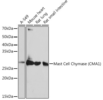
Western blot analysis of various lysates using Mast Cell Chymase (CMA1) (CMA1) Rabbit mAb (CAB11480) at 1:1000 dilution. Secondary antibody: HRP-conjugated Goat anti-Rabbit IgG (H+L) (CABS014) at 1:10000 dilution. Lysates/proteins: 25μg per lane. Blocking buffer: 3% nonfat dry milk in TBST. Detection: ECL Basic Kit (AbGn00020). Exposure time: 10s.
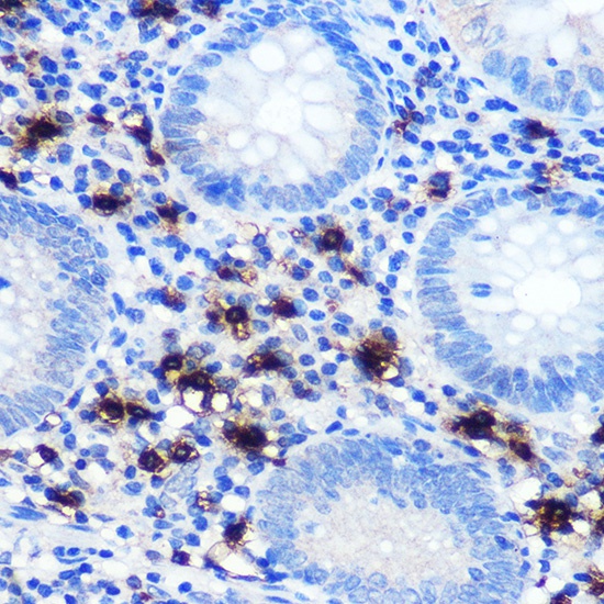
Immunohistochemistry analysis of paraffin-embedded Human appendix using Mast Cell Chymase (CMA1) (CMA1) Rabbit mAb (CAB11480) at dilution of 1:100 (40x lens). Microwave antigen retrieval performed with 0.01M PBS Buffer (pH 7.2) prior to IHC staining.


 ELISA Kit")