The COG1 Antibody (CAB17594) is a high-quality antibody developed for reliable detection and analysis of target proteins. This antibody, produced in rabbits, is highly specific for detecting COG1 in human samples and has been validated for use in Western blot applications. By binding to the COG1 protein, this antibody enables researchers to study its localization and function in various cell types, making it an essential reagent for studies in cell biology and molecular biology.COG1 is a crucial component of the conserved oligomeric Golgi (COG) complex, which is responsible for mediating retrograde vesicular transport between Golgi compartments.
This antibody is validated for use in WB, IHC-P, ELISA applications and has demonstrated reactivity against Human, Mouse, Rat samples.
Product Name:
COG1 Antibody
SKU:
CAB17594
Size:
20μL, 100μL
Reactivity:
Human, Mouse, Rat
Conjugate:
Unconjugated
Immunogen:
Synthetic peptide. This information is considered to be commercially sensitive.
Recommended starting concentration is 1 μg/mL. Please optimize the concentration based on your specific assay requirements.
Synonyms:
LDLB, CDG2G, COG1
Positive Sample:
22Rv1
Cellular Localization:
Golgi Apparatus, Golgi Transport Complex.
Calculated MW:
109kDa
Observed MW:
109kDa
The protein encoded by this gene is one of eight proteins (Cog1-8) which form a Golgi-localized complex (COG) required for normal Golgi morphology and function. It is thought that this protein is required for steps in the normal medial and trans Golgi-associated processing of glycoconjugates and plays a role in the organization of the Golgi-localized complex.
Purification Method
Affinity purification
Gene ID
9382
RRID
AB_2768980
Buffer Information
Store at -20℃. Avoid freeze / thaw cycles. Buffer: PBS with 0.01% thimerosal,50% glycerol,pH7.3.
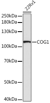
Western blot analysis of lysates from 22Rv1 cells, using COG1 Rabbit pAb (CAB17594) at 1:1000 dilution. Secondary antibody: HRP-conjugated Goat anti-Rabbit IgG (H+L) (CABS014) at 1:10000 dilution. Lysates/proteins: 25μg per lane. Blocking buffer: 3% nonfat dry milk in TBST. Detection: ECL Basic Kit (AbGn00020). Exposure time: 90s.
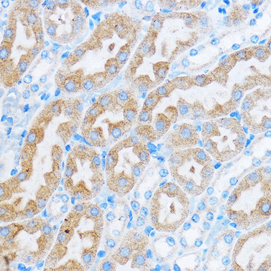
Immunohistochemistry analysis of paraffin-embedded Mouse kidney using COG1 Rabbit pAb (CAB17594) at dilution of 1:50 (40x lens). Microwave antigen retrieval performed with 0.01M PBS Buffer (pH 7.2) prior to IHC staining.
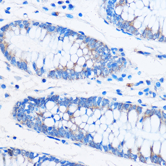
Immunohistochemistry analysis of paraffin-embedded Human colon using COG1 Rabbit pAb (CAB17594) at dilution of 1:50 (40x lens). Microwave antigen retrieval performed with 0.01M PBS Buffer (pH 7.2) prior to IHC staining.