The COLGALT1 Antibody (CAB16571) is a high-quality antibody developed for reliable detection and analysis of target proteins. This antibody, produced in rabbits, exhibits high reactivity with human samples and is validated for use in Western blot applications. By binding specifically to ColGALT1 protein, it enables accurate detection and analysis in a variety of cell types, making it ideal for investigations in fields such as glycobiology, cancer research, and developmental biology.
This antibody is validated for use in WB, IF/ICC, ELISA applications and has demonstrated reactivity against Human, Mouse, Rat samples.
Product Name:
COLGALT1 Antibody
SKU:
CAB16571
Size:
20μL, 100μL
Reactivity:
Human, Mouse, Rat
Conjugate:
Unconjugated
Immunogen:
Recombinant protein (or fragment).This information is considered to be commercially sensitive.
Recommended starting concentration is 1 μg/mL. Please optimize the concentration based on your specific assay requirements.
Synonyms:
BSVD3, GLT25D1, ColGalT 1, COLGALT1
Positive Sample:
Mouse ovary
Cellular Localization:
Endoplasmic Reticulum Lumen.
Calculated MW:
72kDa
Observed MW:
72kDa
The protein encoded by this gene is one of two enzymes that transfers galactose moieties to hydroxylysine residues of collagen and mannose binding lectin. This gene is constitutively expressed and encodes a soluble protein that localizes to the endoplasmic reticulum.
Purification Method
Affinity purification
Gene ID
79709
RRID
AB_2769003
Buffer Information
Store at -20℃. Avoid freeze / thaw cycles. Buffer: PBS containing 50% glycerol, preserved with proclin300 or sodium azide, pH 7.3.
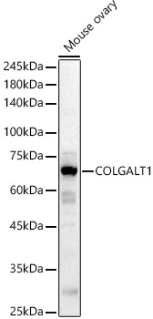
Western blot analysis of lysates from Mouse ovary, using COLGALT1 Rabbit pAb (CAB16571) at 1:1000 dilution. Secondary antibody: HRP-conjugated Goat anti-Rabbit IgG (H+L) (CABS014) at 1:10000 dilution. Lysates/proteins: 25μg per lane. Blocking buffer: 3% nonfat dry milk in TBST. Detection: ECL Basic Kit (AbGn00020). Exposure time: 90s.
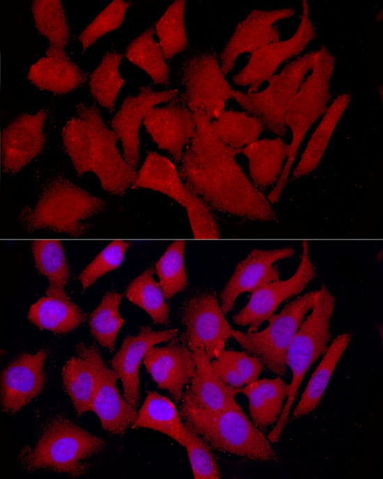
Immunofluorescence analysis of HeLa cells using COLGALT1 Rabbit pAb (CAB16571) at dilution of 1:100 (40x lens). Secondary antibody: Cy3-conjugated Goat anti-Rabbit IgG (H+L) (CABS007) at 1:500 dilution. Blue: DAPI for nuclear staining.
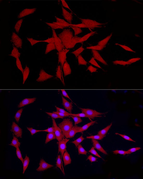
Immunofluorescence analysis of PC-12 cells using COLGALT1 Rabbit pAb (CAB16571) at dilution of 1:100 (40x lens). Secondary antibody: Cy3-conjugated Goat anti-Rabbit IgG (H+L) (CABS007) at 1:500 dilution. Blue: DAPI for nuclear staining.
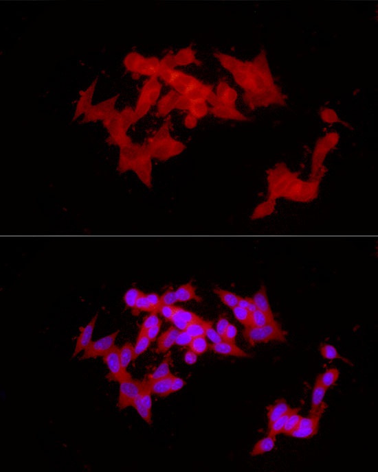
Immunofluorescence analysis of SH-SY5Y cells using COLGALT1 Rabbit pAb (CAB16571) at dilution of 1:100 (40x lens). Secondary antibody: Cy3-conjugated Goat anti-Rabbit IgG (H+L) (CABS007) at 1:500 dilution. Blue: DAPI for nuclear staining.