The C2 Antibody (CAB10186) is a high-quality antibody developed for reliable detection and analysis of target proteins. This antibody, raised in rabbits, specifically targets the Complement C2 protein, a key component of the complement cascade involved in promoting inflammation and opsonization of pathogens.Validated for use in Western blot applications, this antibody is highly reactive with human samples, allowing for the detection and analysis of Complement C2 in various cell types. Its utility in immunology research makes it ideal for studies focusing on the role of the complement system in diseases such as autoimmune disorders, infectious diseases, and inflammatory conditions.
This antibody is validated for use in WB, ELISA applications and has demonstrated reactivity against Human samples.
Product Name:
C2 Antibody
SKU:
CAB10186
Size:
20μL, 100μL
Reactivity:
Human
Conjugate:
Unconjugated
Immunogen:
Synthetic peptide. This information is considered to be commercially sensitive.
Recommended starting concentration is 1 μg/mL. Please optimize the concentration based on your specific assay requirements.
Synonyms:
CO2, ARMD14, C2
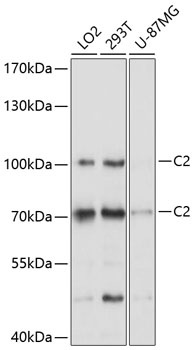
Positive Sample:
LO2, 293T, U-87MG
Cellular Localization:
Secreted.
Calculated MW:
83kDa
Observed MW:
70kDa/110kDa
Component C2 is a serum glycoprotein that functions as part of the classical pathway of the complement system. Activated C1 cleaves C2 into C2a and C2b. The serine proteinase C2a then combines with complement factor 4b to create the C3 or C5 convertase. Deficiency of C2 has been reported to associated with certain autoimmune diseases and SNPs in this gene have been associated with altered susceptibility to age-related macular degeneration. This gene localizes within the class III region of the MHC on the short arm of chromosome 6. Alternative splicing results in multiple transcript variants encoding distinct isoforms. Additional transcript variants have been described in publications but their full-length sequence has not been determined.
Purification Method
Affinity purification
Gene ID
717
RRID
AB_2757707
Buffer Information
Store at -20℃. Avoid freeze / thaw cycles. Buffer: PBS containing 50% glycerol, preserved with proclin300 or sodium azide, pH 7.3.
Western blot analysis of various lysates using C2 Rabbit pAb (CAB10186) at 1:1000 dilution. Secondary antibody: HRP-conjugated Goat anti-Rabbit IgG (H+L) (CABS014) at 1:10000 dilution. Lysates/proteins: 25μg per lane. Blocking buffer: 3% nonfat dry milk in TBST. Detection: ECL Basic Kit (AbGn00020). Exposure time: 5s.