The COPB2 Antibody (CAB7036) is a high-quality antibody developed for reliable detection and analysis of target proteins. This antibody, produced in rabbits, exhibits high specificity and sensitivity towards COPB2 in human samples, making it ideal for Western blot applications. By binding specifically to COPB2, this antibody enables precise detection and analysis of the protein in various cell types, facilitating investigations in molecular biology and cell biology.COPB2, also known as coatomer protein complex subunit beta 2, plays a crucial role in maintaining cellular homeostasis by facilitating the transport of proteins between different cellular compartments.
This antibody is validated for use in WB, IHC-P, IF/ICC, ELISA applications and has demonstrated reactivity against Human, Mouse, Rat samples.
Product Name:
COPB2 Antibody
SKU:
CAB7036
Size:
20μL, 100μL
Reactivity:
Human, Mouse, Rat
Conjugate:
Unconjugated
Immunogen:
Recombinant protein (or fragment).This information is considered to be commercially sensitive.
The Golgi coatomer complex (see MIM 601924) constitutes the coat of nonclathrin-coated vesicles and is essential for Golgi budding and vesicular trafficking. It consists of 7 protein subunits, including COPB2.
Purification Method
Affinity purification
Gene ID
9276
RRID
AB_2767591
Buffer Information
Store at -20℃. Avoid freeze / thaw cycles. Buffer: PBS containing 50% glycerol, preserved with proclin300 or sodium azide,pH7.3.
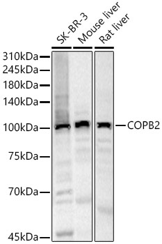
Western blot analysis of various lysates, using COPB2 Rabbit pAb (CAB7036) at 1:500 dilution. Secondary antibody: HRP-conjugated Goat anti-Rabbit IgG (H+L) (CABS014) at 1:10000 dilution. Lysates/proteins: 25μg per lane. Blocking buffer: 3% nonfat dry milk in TBST. Detection: ECL Basic Kit (AbGn00020). Exposure time: 30s.
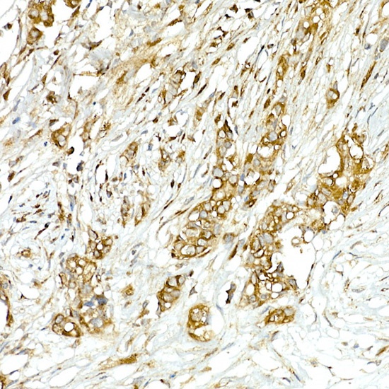
Immunohistochemistry analysis of paraffin-embedded Human breast cancer using COPB2 Rabbit pAb (CAB7036) at dilution of 1:50 (40x lens). High pressure antigen retrieval performed with 0.01M Citrate buffer (pH 6.0) prior to IHC staining.
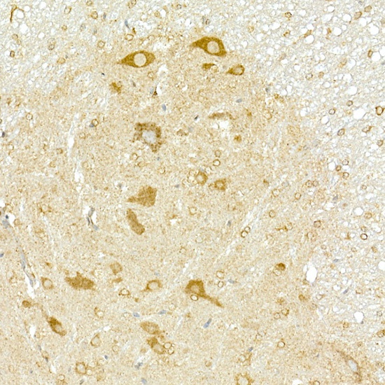
Immunohistochemistry analysis of paraffin-embedded Mouse spinal cord using COPB2 Rabbit pAb (CAB7036) at dilution of 1:50 (40x lens). High pressure antigen retrieval performed with 0.01M Citrate buffer (pH 6.0) prior to IHC staining.
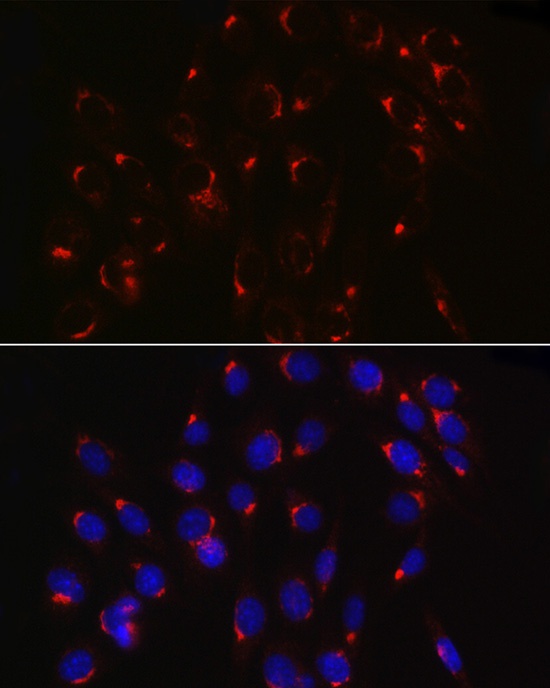
Immunofluorescence analysis of NIH/3T3 cells using COPB2 Rabbit pAb (CAB7036) at dilution of 1:100 (40x lens). Secondary antibody: Cy3-conjugated Goat anti-Rabbit IgG (H+L) (CABS007) at 1:500 dilution. Blue: DAPI for nuclear staining.
Immunofluorescence analysis of U2OS cells using COPB2 Rabbit pAb (CAB7036) at dilution of 1:100 (40x lens). Secondary antibody: Cy3-conjugated Goat anti-Rabbit IgG (H+L) (CABS007) at 1:500 dilution. Blue: DAPI for nuclear staining.