The COPS3 Antibody (CAB7017) is a high-quality antibody developed for reliable detection and analysis of target proteins. The antibody, raised in rabbits, is highly selective for human samples and has been validated for use in Western blot applications. By binding to the COPS3 protein, this antibody enables precise detection and analysis in various cell types, making it an essential tool for studies in molecular biology, cell biology, and cancer research.COPS3, also known as COP9 signalosome subunit 3, is a crucial component of the COP9 signalosome complex that regulates the stability of several proteins involved in cell division and growth.
This antibody is validated for use in WB, IF/ICC, ELISA applications and has demonstrated reactivity against Human, Mouse, Rat samples.
Product Name:
COPS3 Antibody
SKU:
CAB7017
Size:
20μL, 100μL
Reactivity:
Human, Mouse, Rat
Conjugate:
Unconjugated
Immunogen:
Recombinant protein (or fragment).This information is considered to be commercially sensitive.
Recommended starting concentration is 1 μg/mL. Please optimize the concentration based on your specific assay requirements.
Synonyms:
CSN3, SGN3, COPS3
Positive Sample:
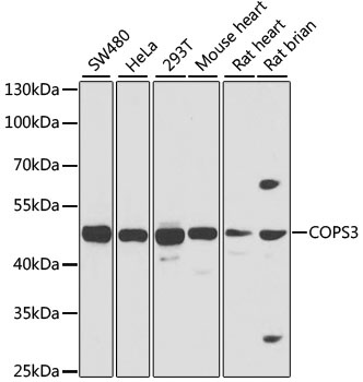
SW480, HeLa, 293T, Mouse heart, Rat heart, Rat brian
Cellular Localization:
Cytoplasm, Nucleus.
Calculated MW:
48kDa
Observed MW:
48kDa
The protein encoded by this gene possesses kinase activity that phosphorylates regulators involved in signal transduction. It phosphorylates I kappa-Balpha, p105, and c-Jun. It acts as a docking site for complex-mediated phosphorylation. The gene is located within the Smith-Magenis syndrome region on chromosome 17. Several transcript variants encoding different isoforms have been found for this gene.
Purification Method
Affinity purification
Gene ID
8533
RRID
AB_2767573
Buffer Information
Store at -20℃. Avoid freeze / thaw cycles. Buffer: PBS containing 50% glycerol, preserved with proclin300 or sodium azide, pH 7.3.
Western blot analysis of various lysates using COPS3 Rabbit pAb (CAB7017) at 1:1000 dilution. Secondary antibody: HRP-conjugated Goat anti-Rabbit IgG (H+L) (CABS014) at 1:10000 dilution. Lysates/proteins: 25μg per lane. Blocking buffer: 3% nonfat dry milk in TBST. Detection: ECL Basic Kit (AbGn00020). Exposure time: 1s.
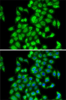
Immunofluorescence analysis of MCF7 cells using COPS3 Rabbit pAb (CAB7017). Secondary antibody: Cy3-conjugated Goat anti-Rabbit IgG (H+L) (CABS007) at 1:500 dilution. Blue: DAPI for nuclear staining.