The COTL1 Antibody (CAB4550) is a high-quality antibody developed for reliable detection and analysis of target proteins. This antibody, raised in rabbits, exhibits high reactivity with human samples and is validated for use in applications such as Western blot and immunohistochemistry.COTL1, also known as Coactosin-like protein 1, plays a crucial role in regulating actin dynamics and cell movement, making it a key player in processes like cell migration and tissue remodeling.
This antibody is validated for use in WB, IHC-P, ELISA applications and has demonstrated reactivity against Human, Mouse, Rat samples.
Product Name:
COTL1 Antibody
SKU:
CAB4550
Size:
20μL, 100μL
Reactivity:
Human, Mouse, Rat
Conjugate:
Unconjugated
Immunogen:
Recombinant protein (or fragment).This information is considered to be commercially sensitive.
This gene encodes one of the numerous actin-binding proteins which regulate the actin cytoskeleton. This protein binds F-actin, and also interacts with 5-lipoxygenase, which is the first committed enzyme in leukotriene biosynthesis. Although this gene has been reported to map to chromosome 17 in the Smith-Magenis syndrome region, the best alignments for this gene are to chromosome 16. The Smith-Magenis syndrome region is the site of two related pseudogenes.
Purification Method
Affinity purification
Gene ID
23406
RRID
AB_2765749
Buffer Information
Store at -20℃. Avoid freeze / thaw cycles. Buffer: PBS containing 50% glycerol, preserved with proclin300 or sodium azide, pH 7.3.
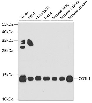
Western blot analysis of various lysates using COTL1 Rabbit pAb (CAB4550) at 1:1000 dilution. Secondary antibody: HRP-conjugated Goat anti-Rabbit IgG (H+L) (CABS014) at 1:10000 dilution. Lysates/proteins: 25μg per lane. Blocking buffer: 3% nonfat dry milk in TBST. Detection: ECL Basic Kit (AbGn00020). Exposure time: 90s.
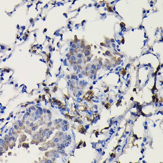
Immunohistochemistry analysis of paraffin-embedded Rat lung using COTL1 Rabbit pAb (CAB4550) at dilution of 1:100 (40x lens). High pressure antigen retrieval performed with 0.01M Citrate buffer (pH 6.0) prior to IHC staining.
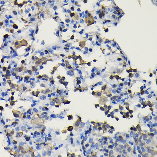
Immunohistochemistry analysis of paraffin-embedded Mouse lung using COTL1 Rabbit pAb (CAB4550) at dilution of 1:100 (40x lens). High pressure antigen retrieval performed with 0.01M Citrate buffer (pH 6.0) prior to IHC staining.