The COX5B Antibody (CAB2640) is a high-quality antibody developed for reliable detection and analysis of target proteins. COX5B is a subunit of cytochrome c oxidase, the final enzyme complex in the mitochondrial electron transport chain. This antibody, raised in rabbits, demonstrates high specificity and sensitivity when detecting COX5B in human samples, making it suitable for Western blot applications.By targeting the COX5B protein, researchers can investigate its involvement in mitochondrial energy production and respiratory function.
This antibody is validated for use in WB, IHC-P, IF/ICC, IP, ELISA applications and has demonstrated reactivity against Human, Mouse, Rat samples.
Product Name:
COX5B Antibody
SKU:
CAB2640
Size:
20μL, 100μL
Reactivity:
Human, Mouse, Rat
Conjugate:
Unconjugated
Immunogen:
Recombinant protein (or fragment).This information is considered to be commercially sensitive.
0.5μg-4μg antibody for 200μg-400μg extracts of whole cells
ELISA
Recommended starting concentration is 1 μg/mL. Please optimize the concentration based on your specific assay requirements.
Synonyms:
COXVB, COX5B
Positive Sample:
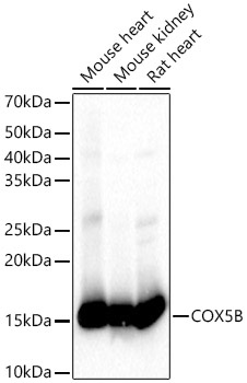
Mouse heart, Mouse kidney, Rat heart , Mouse heart, Mouse kidney, Rat heart
Cellular Localization:
Mitochondrion Inner Membrane.
Calculated MW:
14kDa
Observed MW:
14kDa
Cytochrome C oxidase (COX) is the terminal enzyme of the mitochondrial respiratory chain. It is a multi-subunit enzyme complex that couples the transfer of electrons from cytochrome c to molecular oxygen and contributes to a proton electrochemical gradient across the inner mitochondrial membrane. The complex consists of 13 mitochondrial- and nuclear-encoded subunits. The mitochondrially-encoded subunits perform the electron transfer and proton pumping activities. The functions of the nuclear-encoded subunits are unknown but they may play a role in the regulation and assembly of the complex. This gene encodes the nuclear-encoded subunit Vb of the human mitochondrial respiratory chain enzyme.
Purification Method
Affinity purification
Gene ID
1329
RRID
AB_2764511
Buffer Information
Store at -20℃. Avoid freeze / thaw cycles. Buffer: PBS containing 50% glycerol, preserved with proclin300 or sodium azide, pH 7.3.
Western blot analysis of various lysates using COX5B Rabbit pAb (CAB2640) at 1:1000 dilution. Secondary antibody: HRP-conjugated Goat anti-Rabbit IgG (H+L) (CABS014) at 1:10000 dilution. Lysates / proteins: 25 μg per lane. Blocking buffer: 3 % nonfat dry milk in TBST. Detection: ECL Basic Kit (AbGn00020). Exposure time: 10s.
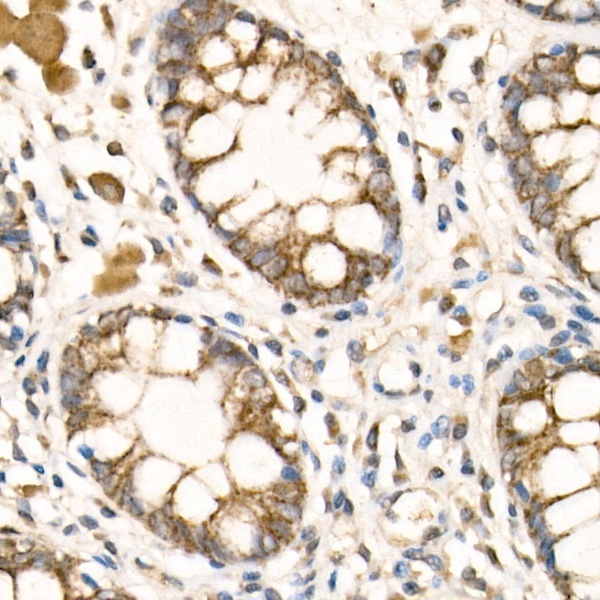
Immunohistochemistry analysis of paraffin-embedded Human colon tissue using COX5B Rabbit pAb (CAB2640) at a dilution of 1:50 (40x lens). High pressure antigen retrieval performed with 0.01M Citrate buffer (pH 6.0) prior to IHC staining.
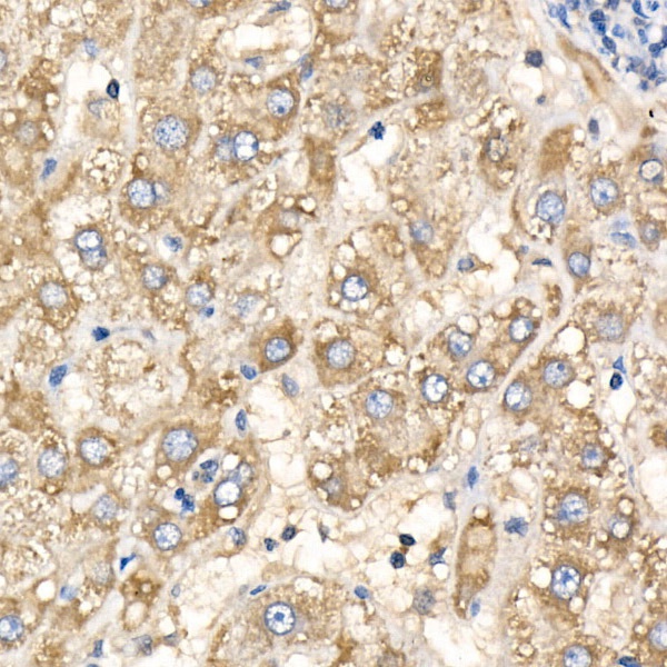
Immunohistochemistry analysis of paraffin-embedded Human liver tissue using COX5B Rabbit pAb (CAB2640) at a dilution of 1:50 (40x lens). High pressure antigen retrieval performed with 0.01M Citrate buffer (pH 6.0) prior to IHC staining.
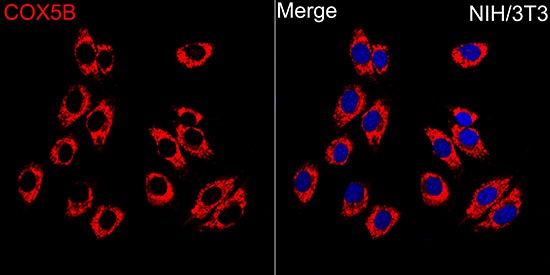
Immunofluorescence analysis of NIH/3T3 cells using COX5B Rabbit pAb (CAB2640) at dilution of 1:100 (40x lens). Secondary antibody: Cy3-conjugated Goat anti-Rabbit IgG (H+L) (CABS007) at 1:500 dilution. Blue: DAPI for nuclear staining.