The CPEB3 Antibody (CAB15402) is a high-quality antibody developed for reliable detection and analysis of target proteins. This antibody, produced in rabbits, exhibits high specificity and sensitivity towards human samples, making it a reliable choice for Western blot applications.CPEB3 is known for its function in controlling the translation of specific mRNAs in response to synaptic activity, contributing to long-lasting changes in neuronal connections. Dysregulation of CPEB3 has been implicated in neurological disorders such as Alzheimer's disease and intellectual disabilities, highlighting its importance in neuronal function and cognitive processes.
This antibody is validated for use in WB, IHC-P, IF/ICC, ELISA applications and has demonstrated reactivity against Human, Mouse, Rat samples.
Product Name:
CPEB3 Antibody
SKU:
CAB15402
Size:
20μL, 100μL
Reactivity:
Human, Mouse, Rat
Conjugate:
Unconjugated
Immunogen:
Recombinant protein (or fragment).This information is considered to be commercially sensitive.
Enables mRNA 3'-UTR binding activity and translation factor activity, RNA binding. Involved in cellular response to amino acid stimulus; negative regulation of transcription by RNA polymerase II; and positive regulation of mRNA catabolic process. Located in several cellular components, including cytosol; midbody; and nucleoplasm. Part of CCR4-NOT complex.
Purification Method
Affinity purification
Gene ID
22849
RRID
AB_2762309
Buffer Information
Store at -20℃. Avoid freeze / thaw cycles. Buffer: PBS containing 50% glycerol, preserved with proclin300 or sodium azide, pH 7.3.
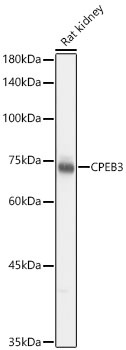
Western blot analysis of lysates from Rat kidney using CPEB3 Rabbit pAb (CAB15402) at 1:1000 dilution. Secondary antibody: HRP-conjugated Goat anti-Rabbit IgG (H+L) (CABS014) at 1:10000 dilution. Lysates/proteins: 25 μg per lane. Blocking buffer: 3% nonfat dry milk in TBST. Detection: ECL Basic Kit (AbGn00020). Exposure time: 1s.
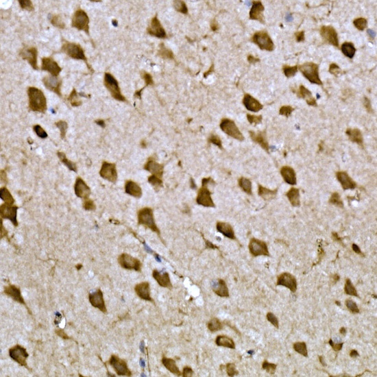
Immunohistochemistry analysis of paraffin-embedded Mouse brain tissue using CPEB3 Rabbit pAb (CAB15402) at a dilution of 1:300 (40x lens). High pressure antigen retrieval performed with 0.01M Citrate buffer (pH 6.0) prior to IHC staining.
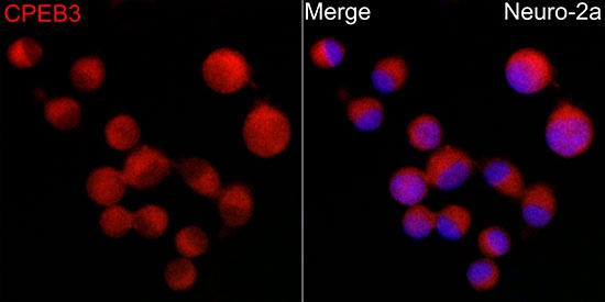
Immunofluorescence analysis of Neuro-2a cells using CPEB3 Rabbit pAb (CAB15402) at dilution of 1:200 (40x lens). Secondary antibody: Cy3-conjugated Goat anti-Rabbit IgG (H+L) (CABS007) at 1:500 dilution. Blue: DAPI for nuclear staining.