The HSPE1/HSP10/CPN10 Monoclonal Antibody (CAB5580) is a high-quality antibody developed for reliable detection and analysis of target proteins. This gene encodes a major heat shock protein which functions as a chaperonin. Its structure consists of a heptameric ring which binds to another heat shock protein in order to form a symmetric, functional heterodimer which enhances protein folding in an ATP-dependent manner. This gene and its co-chaperonin, HSPD1, are arranged in a head-to-head orientation on chromosome 2. Naturally occurring read-through transcription occurs between this locus and the neighboring locus MOBKL3.
This antibody is validated for use in WB, IHC-P, ELISA applications and has demonstrated reactivity against Human, Mouse, Rat samples.
Product Name:
HSPE1/HSP10/CPN10 Monoclonal Antibody
SKU:
CAB5580
Size:
100μL, 20μL
Reactivity:
Human, Mouse, Rat
Clone Number:
ARC1411
Conjugate:
Unconjugated
Immunogen:
Recombinant protein (or fragment).This information is considered to be commercially sensitive.
Tested Applications:
WBIHC-PELISA
Recommended Dilution:
WB
1:1000 - 1:6000
IHC-P
1:100 - 1:500
ELISA
Recommended starting concentration is 1 μg/mL. Please optimize the concentration based on your specific assay requirements.
Synonyms:
EPF, CPN10, GROES, HSP10, HSPE1/HSP10/CPN10
Positive Sample:
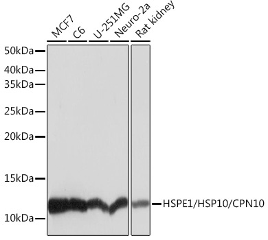
MCF7, C6, U-251MG, Neuro-2a, Rat kidney
Cellular Localization:
Mitochondrion Matrix.
Calculated MW:
11kDa
Observed MW:
11kDa
This gene encodes a major heat shock protein which functions as a chaperonin. Its structure consists of a heptameric ring which binds to another heat shock protein in order to form a symmetric, functional heterodimer which enhances protein folding in an ATP-dependent manner. This gene and its co-chaperonin, HSPD1, are arranged in a head-to-head orientation on chromosome 2. Naturally occurring read-through transcription occurs between this locus and the neighboring locus MOBKL3.
Purification Method
Affinity purification
Gene ID
3336
RRID
AB_2863510
Buffer Information
Store at -20℃. Avoid freeze / thaw cycles. Buffer: PBS containing 50% glycerol and 0.05% BSA, preserved with proclin300 or sodium azide, pH 7.3.
Western blot analysis of various lysates using HSPE1/HSP10/HSPE1/HSP10/CPN10 Rabbit mAb (CAB5580) at 1:1000 dilution. Secondary antibody: HRP-conjugated Goat anti-Rabbit IgG (H+L) (AS014) at 1:10000 dilution. Lysates/proteins: 25μg per lane. Blocking buffer: 3% nonfat dry milk in TBST. Detection: ECL Basic Kit (AbGn00020). Exposure time: 1s.
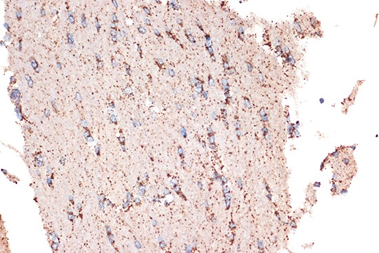
Immunohistochemistry analysis of paraffin-embedded Rat brain using HSPE1/HSP10/HSPE1/HSP10/CPN10 Rabbit mAb (CAB5580) at dilution of 1:100 (40x lens). Microwave antigen retrieval performed with 0.01M Tris/EDTA Buffer (pH 9.0) prior to IHC staining.
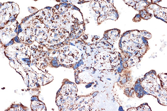
Immunohistochemistry analysis of paraffin-embedded Human placenta using HSPE1/HSP10/HSPE1/HSP10/CPN10 Rabbit mAb (CAB5580) at dilution of 1:100 (40x lens). Microwave antigen retrieval performed with 0.01M Tris/EDTA Buffer (pH 9.0) prior to IHC staining.
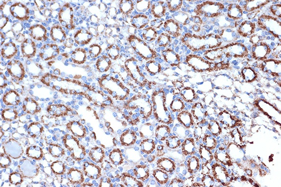
Immunohistochemistry analysis of paraffin-embedded Mouse kidney using HSPE1/HSP10/HSPE1/HSP10/CPN10 Rabbit mAb (CAB5580) at dilution of 1:100 (40x lens). Microwave antigen retrieval performed with 0.01M Tris/EDTA Buffer (pH 9.0) prior to IHC staining.
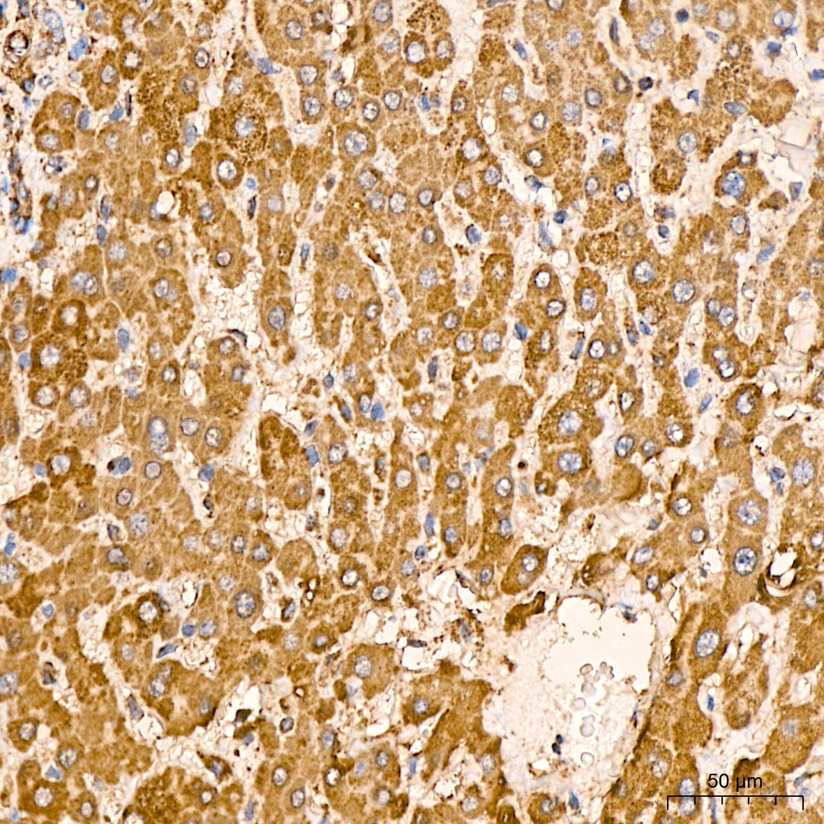
Immunohistochemistry analysis of paraffin-embedded Human liver tissue using HSPE1/HSP10/CPN10 Rabbit mAb (CAB5580) at a dilution of 1:400 (40x lens). High pressure antigen retrieval performed with 0.01M Citrate buffer (pH 6.0) prior to IHC staining.
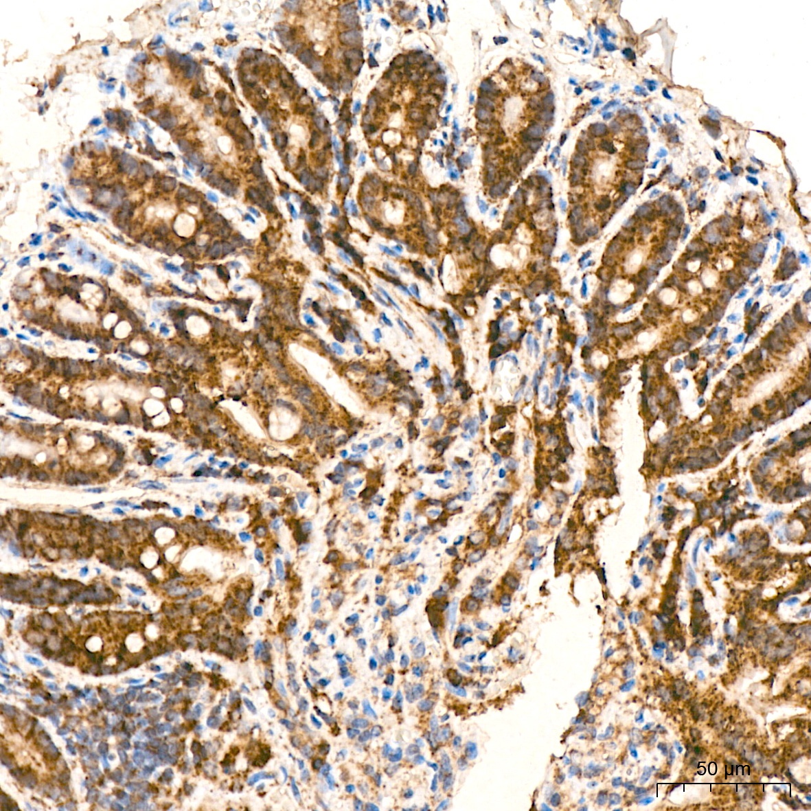
Immunohistochemistry analysis of paraffin-embedded Mouse colon tissue using HSPE1/HSP10/CPN10 Rabbit mAb (CAB5580) at a dilution of 1:400 (40x lens). High pressure antigen retrieval performed with 0.01M Citrate buffer (pH 6.0) prior to IHC staining.


")

