The CRMP1 Antibody (CAB2705) is a high-quality antibody developed for reliable detection and analysis of target proteins. This gene encodes a member of a family of cytosolic phosphoproteins expressed exclusively in the nervous system. The encoded protein is thought to be a part of the semaphorin signal transduction pathway implicated in semaphorin-induced growth cone collapse during neural development. Alternative splicing results in multiple transcript variants.
This antibody is validated for use in WB, IF/ICC, ELISA applications and has demonstrated reactivity against Human, Mouse, Rat samples.
Product Name:
CRMP1 Antibody
SKU:
CAB2705
Size:
100μL, 20μL
Reactivity:
Human, Mouse, Rat
Conjugate:
Unconjugated
Immunogen:
Recombinant protein (or fragment).This information is considered to be commercially sensitive.
Tested Applications:
WBIF/ICCELISA
Recommended Dilution:
WB
1:500 - 1:1000
IF/ICC
1:100 - 1:500
ELISA
Recommended starting concentration is 1 μg/mL. Please optimize the concentration based on your specific assay requirements.
Synonyms:
DRP1, DRP-1, CRMP-1, DPYSL1, ULIP-3, CRMP1
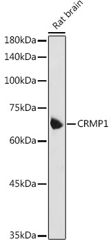
Positive Sample:
Rat brain
Cellular Localization:
Cytoplasm, Centrosome, Cytoskeleton, Microtubule Organizing Center, Spindle.
Calculated MW:
62kDa
Observed MW:
62kDa
This gene encodes a member of a family of cytosolic phosphoproteins expressed exclusively in the nervous system. The encoded protein is thought to be a part of the semaphorin signal transduction pathway implicated in semaphorin-induced growth cone collapse during neural development. Alternative splicing results in multiple transcript variants.
Purification Method
Affinity purification
Gene ID
1400
RRID
AB_2764560
Buffer Information
Store at -20℃. Avoid freeze / thaw cycles. Buffer: PBS containing 50% glycerol, preserved with proclin300 or sodium azide, pH 7.3.
Western blot analysis of lysates from Rat brain, using CRMP1 Rabbit pAb (CAB2705) at 1:1000 dilution. Secondary antibody: HRP-conjugated Goat anti-Rabbit IgG (H+L) (AS014) at 1:10000 dilution. Lysates/proteins: 25μg per lane. Blocking buffer: 3% nonfat dry milk in TBST. Detection: ECL Basic Kit (AbGn00020). Exposure time: 5s.
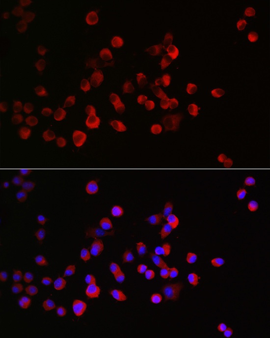
Immunofluorescence analysis of Neuro-2a cells using CRMP1 Rabbit pAb (CAB2705) at dilution of 1:300 (40x lens). Secondary antibody: Cy3-conjugated Goat anti-Rabbit IgG (H+L) (AS007) at 1:500 dilution. Blue: DAPI for nuclear staining.