The CRMP1 Antibody (CAB2705) is a high-quality antibody developed for reliable detection and analysis of target proteins. This antibody, developed using rabbit immunoglobulins, is highly reactive with human samples and has been validated for use in Western blot applications. By binding to the CRMP1 protein, this antibody allows for the detection and analysis of CRMP1 in various cell types, making it an ideal tool for studies in neuroscience, cancer research, and other fields.CRMP1 is a cytosolic protein that is involved in axon guidance and neurite outgrowth, playing a crucial role in neuronal development and function.
This antibody is validated for use in WB, IF/ICC, ELISA applications and has demonstrated reactivity against Human, Mouse, Rat samples.
Product Name:
CRMP1 Antibody
SKU:
CAB2705
Size:
20μL, 100μL
Reactivity:
Human, Mouse, Rat
Conjugate:
Unconjugated
Immunogen:
Recombinant protein (or fragment).This information is considered to be commercially sensitive.
Recommended starting concentration is 1 μg/mL. Please optimize the concentration based on your specific assay requirements.
Synonyms:
DRP1, DRP-1, CRMP-1, DPYSL1, ULIP-3, CRMP1
Positive Sample:
Rat brain
Cellular Localization:
Cytoplasm, Centrosome, Cytoskeleton, Microtubule Organizing Center, Spindle.
Calculated MW:
62kDa
Observed MW:
62kDa
This gene encodes a member of a family of cytosolic phosphoproteins expressed exclusively in the nervous system. The encoded protein is thought to be a part of the semaphorin signal transduction pathway implicated in semaphorin-induced growth cone collapse during neural development. Alternative splicing results in multiple transcript variants.
Purification Method
Affinity purification
Gene ID
1400
RRID
AB_2764560
Buffer Information
Store at -20℃. Avoid freeze / thaw cycles. Buffer: PBS containing 50% glycerol, preserved with proclin300 or sodium azide, pH 7.3.
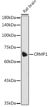
Western blot analysis of lysates from Rat brain, using CRMP1 Rabbit pAb (CAB2705) at 1:1000 dilution. Secondary antibody: HRP-conjugated Goat anti-Rabbit IgG (H+L) (CABS014) at 1:10000 dilution. Lysates/proteins: 25μg per lane. Blocking buffer: 3% nonfat dry milk in TBST. Detection: ECL Basic Kit (AbGn00020). Exposure time: 5s.
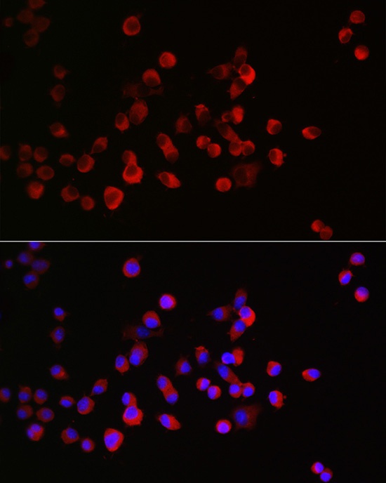
Immunofluorescence analysis of Neuro-2a cells using CRMP1 Rabbit pAb (CAB2705) at dilution of 1:300 (40x lens). Secondary antibody: Cy3-conjugated Goat anti-Rabbit IgG (H+L) (CABS007) at 1:500 dilution. Blue: DAPI for nuclear staining.