The CRY1 Antibody (CAB13662) is a high-quality antibody developed for reliable detection and analysis of target proteins. This antibody, produced in rabbits, exhibits high reactivity with human samples and is validated for use in Western blot applications. By binding specifically to the CRY1 protein, this antibody enables accurate detection and analysis in various cell types, making it an ideal choice for studies in chronobiology, neurology, and metabolism research.CRY1 is a critical component of the circadian clock system, responsible for regulating daily rhythms in processes such as sleep-wake cycles, hormone secretion, and metabolism.
This antibody is validated for use in WB, IHC-P, ELISA applications and has demonstrated reactivity against Human, Mouse, Rat samples.
Product Name:
CRY1 Antibody
SKU:
CAB13662
Size:
20μL, 100μL
Reactivity:
Human, Mouse, Rat
Conjugate:
Unconjugated
Immunogen:
Recombinant protein (or fragment).This information is considered to be commercially sensitive.
Recommended starting concentration is 1 μg/mL. Please optimize the concentration based on your specific assay requirements.
Synonyms:
DSPD, PHLL1, CRY1
Positive Sample:
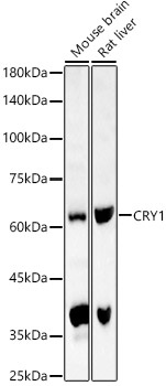
Mouse brain, Rat liver
Cellular Localization:
Cytoplasm, Nucleus.
Calculated MW:
66kDa
Observed MW:
66kDa
This gene encodes a flavin adenine dinucleotide-binding protein that is a key component of the circadian core oscillator complex, which regulates the circadian clock. This gene is upregulated by CLOCK/ARNTL heterodimers but then represses this upregulation in a feedback loop using PER/CRY heterodimers to interact with CLOCK/ARNTL. Polymorphisms in this gene have been associated with altered sleep patterns. The encoded protein is widely conserved across plants and animals. Loss of the related gene in mouse results in a shortened circadian cycle in complete darkness.
Purification Method
Affinity purification
Gene ID
1407
RRID
AB_2760523
Buffer Information
Store at -20℃. Avoid freeze / thaw cycles. Buffer: PBS containing 50% glycerol, preserved with proclin300 or sodium azide, pH 7.3.
Western blot analysis of various lysates, using CRY1 Rabbit pAb (CAB13662) at 1:800 dilution. Secondary antibody: HRP-conjugated Goat anti-Rabbit IgG (H+L) (CABS014) at 1:10000 dilution. Lysates/proteins: 25μg per lane. Blocking buffer: 3% nonfat dry milk in TBST. Detection: ECL Basic Kit (AbGn00020). Exposure time: 30s.
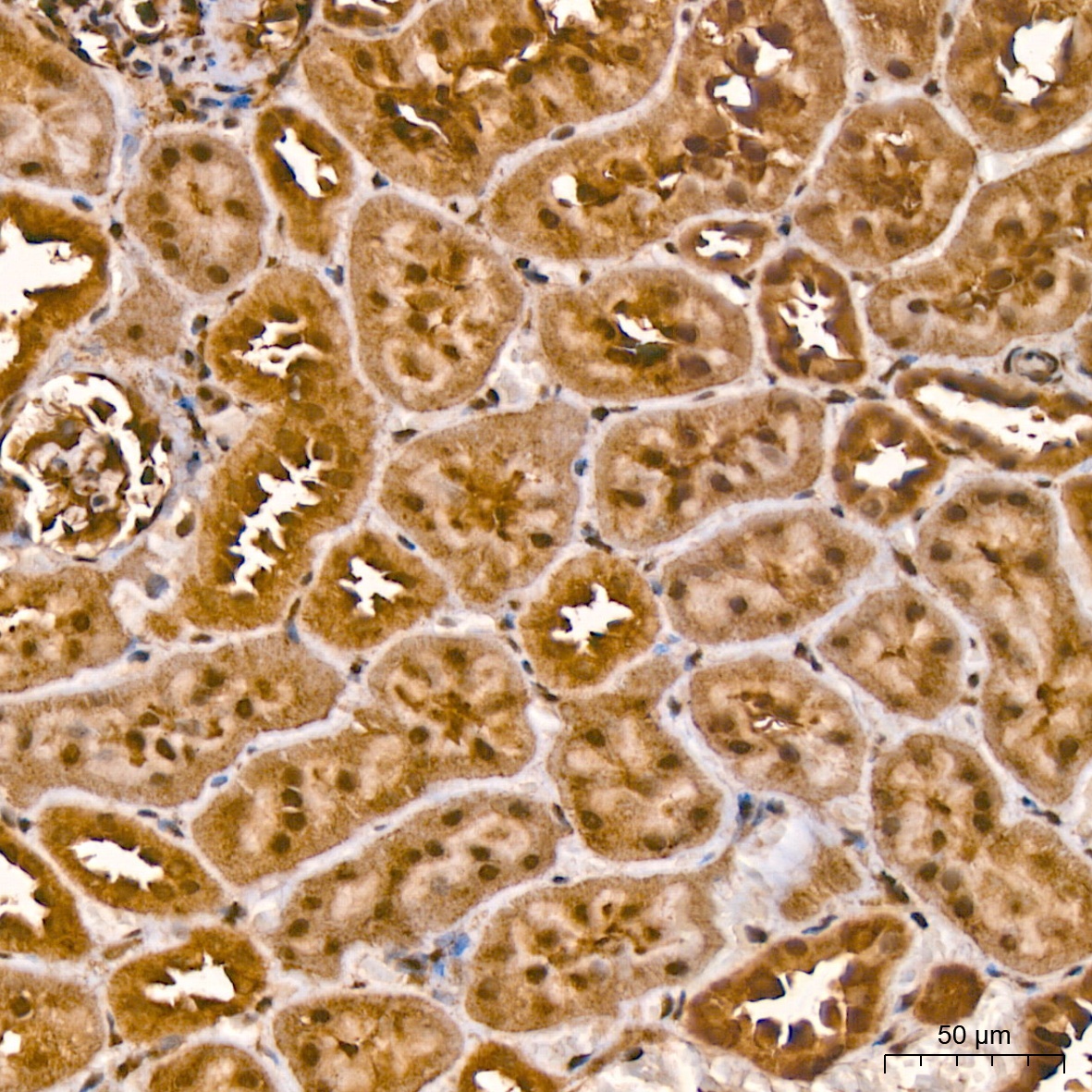
Immunohistochemistry analysis of paraffin-embedded Rat kidney tissue using CRY1 Rabbit pAb (CAB13662) at a dilution of 1:200 (40x lens). High pressure antigen retrieval was performed with 0.01 M citrate buffer (pH 6.0) prior to IHC staining.
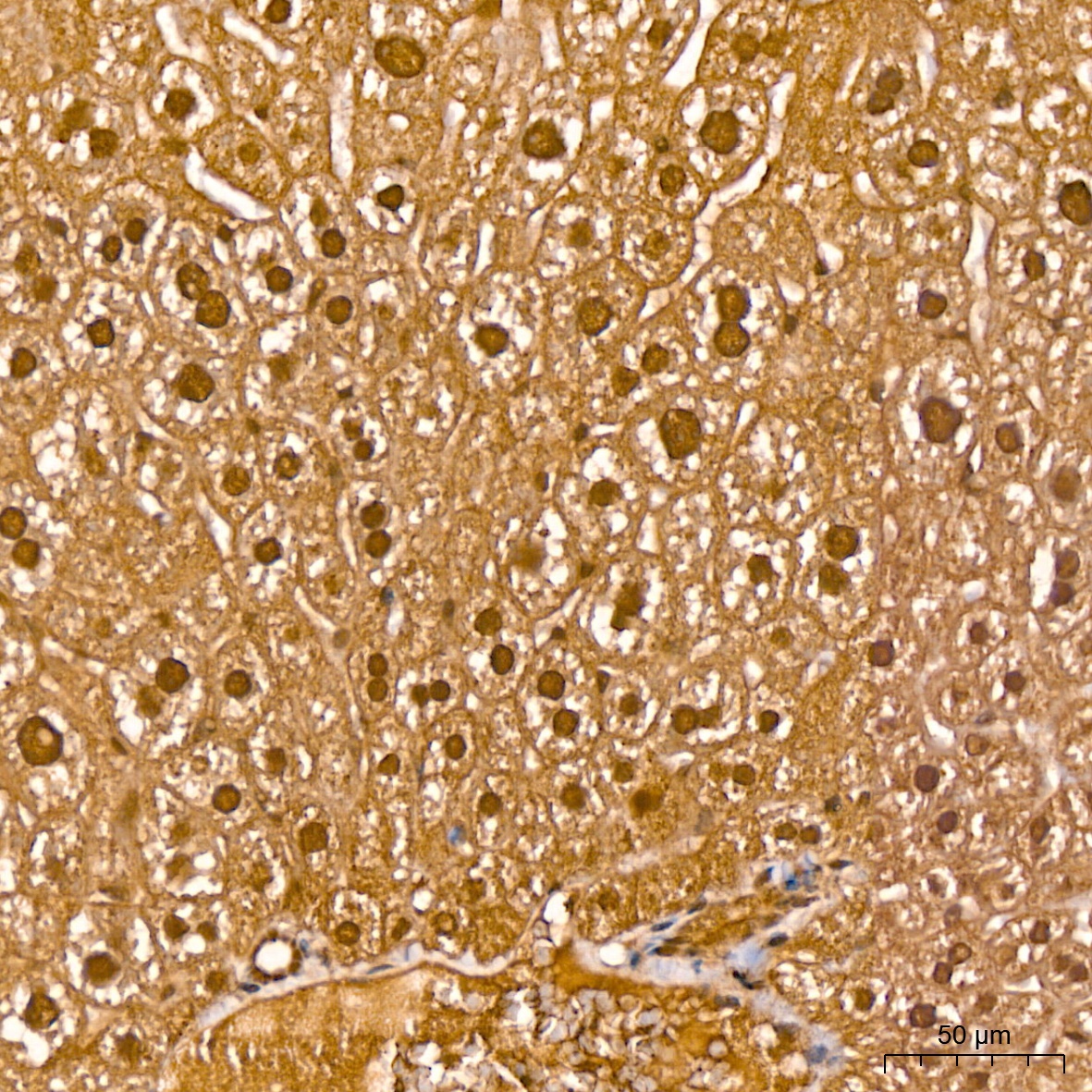
Immunohistochemistry analysis of paraffin-embedded Mouse liver tissue using CRY1 Rabbit pAb (CAB13662) at a dilution of 1:200 (40x lens). High pressure antigen retrieval was performed with 0.01 M citrate buffer (pH 6.0) prior to IHC staining.