The CSF1R Antibody (CAB3019) is a high-quality antibody developed for reliable detection and analysis of target proteins. This antibody, produced through the immunization of rabbits, exhibits high reactivity with human samples and has been validated for use in Western blot applications. By binding specifically to the CSF1R protein, this antibody enables accurate detection and analysis in a variety of cell types, making it an excellent tool for studies in immunology and cancer research.CSF1R, also known as colony-stimulating factor 1 receptor, is a crucial player in immune system regulation and is implicated in various diseases, including cancer and inflammatory conditions.
This antibody is validated for use in WB, IF/ICC, ELISA, IF-P applications and has demonstrated reactivity against Human, Mouse, Rat samples.
Product Name:
CSF1R Antibody
SKU:
CAB3019
Size:
20μL, 100μL
Reactivity:
Human, Mouse, Rat
Conjugate:
Unconjugated
Immunogen:
Synthetic peptide. This information is considered to be commercially sensitive.
Cell Membrane, Single-Pass Type I Membrane Protein.
Calculated MW:
108kDa
Observed MW:
150-190kDa
The protein encoded by this gene is the receptor for colony stimulating factor 1, a cytokine which controls the production, differentiation, and function of macrophages. This receptor mediates most if not all of the biological effects of this cytokine. Ligand binding activates the receptor kinase through a process of oligomerization and transphosphorylation. The encoded protein is a tyrosine kinase transmembrane receptor and member of the CSF1/PDGF receptor family of tyrosine-protein kinases. Mutations in this gene have been associated with a predisposition to myeloid malignancy. The first intron of this gene contains a transcriptionally inactive ribosomal protein L7 processed pseudogene oriented in the opposite direction. Alternative splicing results in multiple transcript variants. Expression of a splice variant from an LTR promoter has been found in Hodgkin lymphoma (HL), HL cell lines and anaplastic large cell lymphoma.
Purification Method
Affinity purification
Gene ID
1436
RRID
AB_2764828
Buffer Information
Store at -20℃. Avoid freeze / thaw cycles. Buffer: PBS containing 50% glycerol, preserved with proclin300 or sodium azide, pH 7.3.
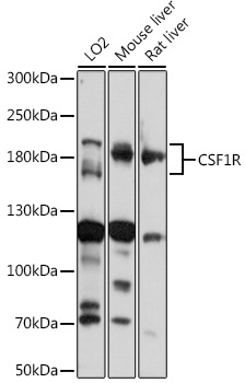
Western blot analysis of various lysates using CD115/CSF-1R Rabbit pAb (CAB3019) at 1:1000 dilution. Secondary antibody: HRP-conjugated Goat anti-Rabbit IgG (H+L) (CABS014) at 1:10000 dilution. Lysates/proteins: 25μg per lane. Blocking buffer: 3% nonfat dry milk in TBST. Detection: ECL Basic Kit (AbGn00020). Exposure time: 5s.
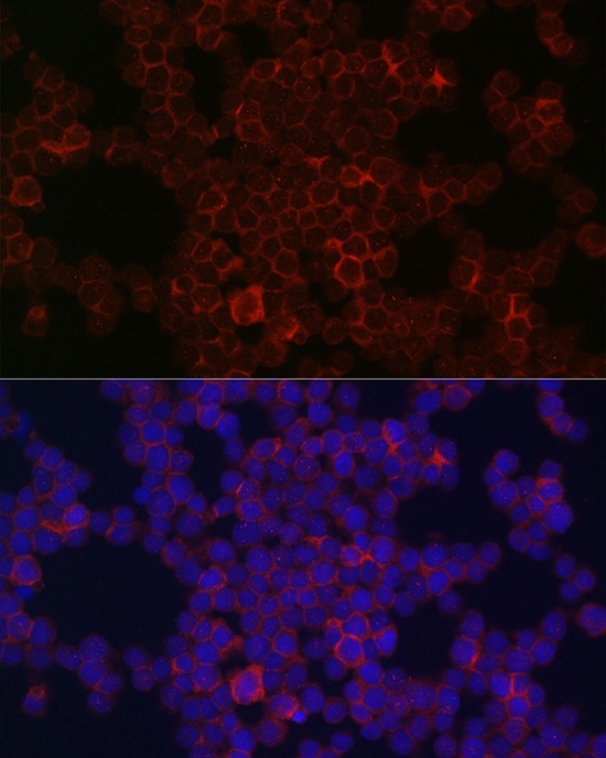
Immunofluorescence analysis of THP-1 cells using CD115/CSF-1R Rabbit pAb (CAB3019) at dilution of 1:100 (40x lens). Secondary antibody: Cy3-conjugated Goat anti-Rabbit IgG (H+L) (CABS007) at 1:500 dilution. Blue: DAPI for nuclear staining.
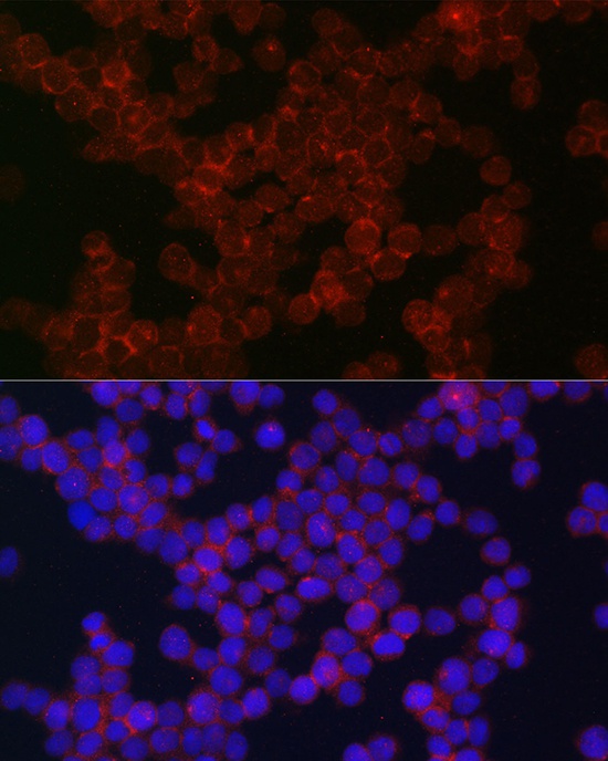
Immunofluorescence analysis of THP-1 cells using CD115/CSF-1R Rabbit pAb (CAB3019) at dilution of 1:100 (40x lens). Secondary antibody: Cy3-conjugated Goat anti-Rabbit IgG (H+L) (CABS007) at 1:500 dilution. Blue: DAPI for nuclear staining.
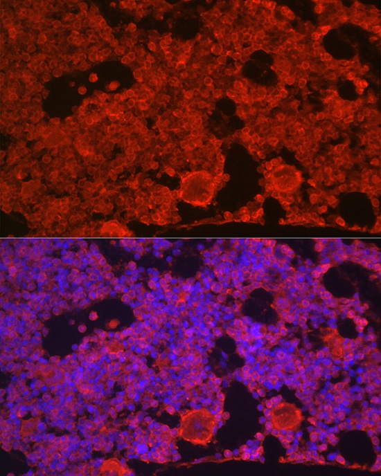
Immunofluorescence analysis of paraffin-embedded mouse bone marrow using CD115/CSF-1R Rabbit pAb (CAB3019) at dilution of 1:100 (40x lens). Secondary antibody: Cy3-conjugated Goat anti-Rabbit IgG (H+L) (CABS007) at 1:500 dilution. Blue: DAPI for nuclear staining.