The CSF2 Antibody (CAB6127) is a high-quality antibody developed for reliable detection and analysis of target proteins. This antibody, produced in rabbits, exhibits high reactivity with human samples and has been validated for use in Western blot assays. By binding specifically to the CSF2 protein, this antibody allows for accurate detection and analysis in a variety of cell types, making it an ideal choice for studies in immunology and cancer research.CSF2, also known as Granulocyte-Macrophage Colony-Stimulating Factor (GM-CSF), is essential for the development and function of various immune cells, including granulocytes, macrophages, and dendritic cells.
This antibody is validated for use in WB, IF/ICC, ELISA applications and has demonstrated reactivity against Human, Mouse, Rat samples.
Product Name:
CSF2 Antibody
SKU:
CAB6127
Size:
20μL, 100μL
Reactivity:
Human, Mouse, Rat
Conjugate:
Unconjugated
Immunogen:
Synthetic peptide. This information is considered to be commercially sensitive.
Sequence:
ARSP SPST QPWE HVNA I
Tested Applications:
WBIF/ICCELISA
Recommended Dilution:
WB
1:500 - 1:2000
IF/ICC
1:50 - 1:200
ELISA
Recommended starting concentration is 1 μg/mL. Please optimize the concentration based on your specific assay requirements.
Synonyms:
CSF, GMCSF, CSF2
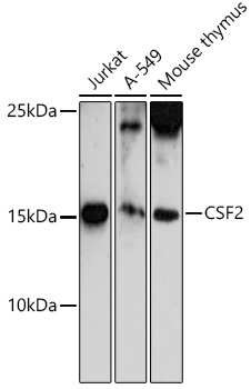
Positive Sample:
Jurkat, A-549, Mouse thymus
Cellular Localization:
Secreted.
Calculated MW:
16kDa
Observed MW:
16kDa
The protein encoded by this gene is a cytokine that controls the production, differentiation, and function of granulocytes and macrophages. The active form of the protein is found extracellularly as a homodimer. This gene has been localized to a cluster of related genes at chromosome region 5q31, which is known to be associated with interstitial deletions in the 5q- syndrome and acute myelogenous leukemia. Other genes in the cluster include those encoding interleukins 4, 5, and 13. This gene plays a role in promoting tissue inflammation. Elevated levels of cytokines, including the one produced by this gene, have been detected in SARS-CoV-2 infected patients that develop acute respiratory distress syndrome. Mice deficient in this gene or its receptor develop pulmonary alveolar proteinosis.
Purification Method
Affinity purification
Gene ID
1437
RRID
AB_2766760
Buffer Information
Store at -20℃. Avoid freeze / thaw cycles. Buffer: PBS containing 50% glycerol, preserved with proclin300 or sodium azide, pH 7.3.
Western blot analysis of various lysates using CSF2 Rabbit pAb (CAB6127) at 1:500 dilution. Secondary antibody: HRP-conjugated Goat anti-Rabbit IgG (H+L) (CABS014) at 1:10000 dilution. Lysates/proteins: 25μg per lane. Blocking buffer: 3% nonfat dry milk in TBST. Detection: ECL Basic Kit (AbGn00020). Exposure time: 120s.
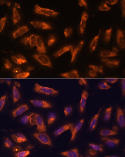
Immunofluorescence analysis of C6 cells using CSF2 Rabbit pAb (CAB6127) at dilution of 1:100. Secondary antibody: Cy3-conjugated Goat anti-Rabbit IgG (H+L) (CABS007) at 1:500 dilution. Blue: DAPI for nuclear staining.
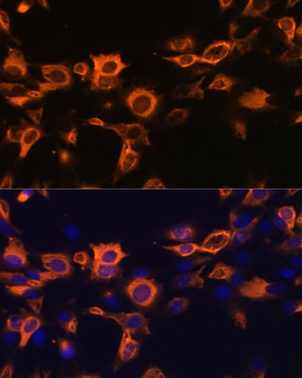
Immunofluorescence analysis of U-2 OS cells using CSF2 Rabbit pAb (CAB6127) at dilution of 1:100. Secondary antibody: Cy3-conjugated Goat anti-Rabbit IgG (H+L) (CABS007) at 1:500 dilution. Blue: DAPI for nuclear staining.