The Cy3-conjugated Goat anti-Mouse IgG (H+L) (CABS008) is a high-quality antibody developed for reliable detection and analysis of target proteins. This antibody is ideal for visualizing and detecting mouse IgG in various biological samples, allowing for precise identification and localization of target proteins.With a bright and stable Cy3 fluorescent label, this antibody offers exceptional performance in fluorescent microscopy and imaging studies. Its conjugation to Cy3 allows for easy and rapid visualization of mouse IgG in tissue sections, cells, or other samples, making it a valuable tool for researchers studying immune responses, antibody localization, and protein expression patterns.
This antibody is validated for use in IF/ICC, FC applications and has demonstrated reactivity against Mouse samples.
Product Name:
Cy3-conjugated Goat anti-Mouse IgG (H+L)
SKU:
CABS008
Size:
100μL
Reactivity:
Mouse
Conjugate:
Cy3. Ex:548nm. Em:562nm.
Immunogen:
This information is considered to be commercially sensitive.
Tested Applications:
IF/ICCFC
Recommended Dilution:
IF/ICC
1:50 - 1:200
FC
1:100 - 1:800
Secondary antibodies are affinity-purified antibodies which will work with target-specific primary antibody in the detection, sorting or purification of its specified target. Secondary antibodies offer increased versatility enabling users to use many detection systems (e.g. HRP, AP, fluorescence). They can also provide greater sensitivity through signal amplification as multiple secondary antibodies . Most commonly, secondary antibodies are generated by immunizing the host animal (different from host species of primary antibody) with a pooled population of normal immunoglobulins from the host species of primary antibody and can be further purified and modified (i.e. antibody fragmentation, label conjugation, etc.) to ensure well-characterized specificity to corresponding normal immunoglobulins.
Purification Method
Affinity purification
RRID
AB_2769088
Buffer Information
Store at -20℃. Avoid freeze / thaw cycles. Buffer: PBS with 0.025% Sodium Azide,0.75% BSA,50% glycerol,pH7.3.
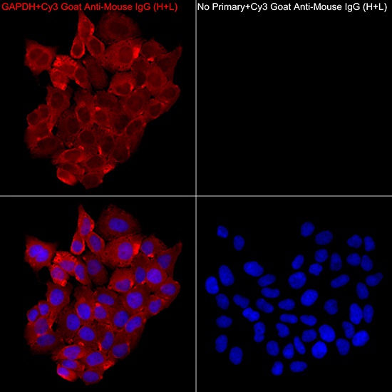
Immunofluorescence analysis of HeLa cells using GAPDH Mouse mAb (AC033, dilution 1:100) followed by a further incubation with Cy3 Goat Anti-Mouse IgG (H+L)(CABS008, dilution 1:200) (Red). DAPI was used for nuclear staining (Blue). Objective: 40x.
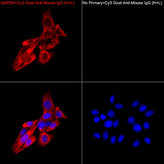
Immunofluorescence analysis of NIH/3T3 cells using GAPDH Mouse mAb (AC033, dilution 1:100) followed by a further incubation with Cy3 Goat Anti-Mouse IgG (H+L)(CABS008, dilution 1:200) (Red). DAPI was used for nuclear staining (Blue). Objective: 40x.
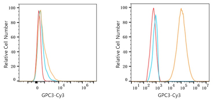
Flow cytometry: 1X10^6 K-562 cells (negative control,left) and Hep G2 cells (right) were surface-stained with Mouse Anti-Human GPC3 mAb (4μg/mL,orange line) or secondary antibody only (blue line). Non-fluorescently stained HepG2 and K-562 cells were used as blank control (red line). Cy3 Goat Anti-Mouse IgG (H+L) (CABS008, 1:200) was used as a secondary antibody.
 (CABS008)")
 (CABS008)")
 (CABS007)")
 (CABS001)")
 (CABS003)")
 (CABS026)")
 (CABS028)")