The Cyclin H Monoclonal Antibody (CAB4076) is a high-quality antibody developed for reliable detection and analysis of target proteins. The protein encoded by this gene belongs to the highly conserved cyclin family, whose members are characterized by a dramatic periodicity in protein abundance through the cell cycle. Cyclins function as regulators of CDK kinases. Different cyclins exhibit distinct expression and degradation patterns which contribute to the temporal coordination of each mitotic event. This cyclin forms a complex with CDK7 kinase and ring finger protein MAT1. The kinase complex is able to phosphorylate CDK2 and CDC2 kinases, thus functions as a CDK-activating kinase (CAK). This cyclin and its kinase partner are components of TFIIH, as well as RNA polymerase II protein complexes. They participate in two different transcriptional regulation processes, suggesting an important link between basal transcription control and the cell cycle machinery. A pseudogene of this gene is found on chromosome 4. Alternate splicing results in multiple transcript variants.
This antibody is validated for use in WB, IHC-P, IF/ICC, ELISA applications and has demonstrated reactivity against Human, Mouse samples.
Product Name:
Cyclin H Monoclonal Antibody
SKU:
CAB4076
Size:
100μL, 20μL
Reactivity:
Human, Mouse
Clone Number:
ARC0893
Conjugate:
Unconjugated
Immunogen:
Synthetic peptide. This information is considered to be commercially sensitive.
Tested Applications:
WBIHC-PIF/ICCELISA
Recommended Dilution:
WB
1:500 - 1:1000
IHC-P
1:50 - 1:200
IF/ICC
1:50 - 1:200
ELISA
Recommended starting concentration is 1 μg/mL. Please optimize the concentration based on your specific assay requirements.
Synonyms:
CAK, p34, p37, CycH, Cyclin H
Positive Sample:
HeLa, Hep G2, Jurkat, Mouse testis
Cellular Localization:
Nucleus.
Calculated MW:
38kDa
Observed MW:
36kDa
The protein encoded by this gene belongs to the highly conserved cyclin family, whose members are characterized by a dramatic periodicity in protein abundance through the cell cycle. Cyclins function as regulators of CDK kinases. Different cyclins exhibit distinct expression and degradation patterns which contribute to the temporal coordination of each mitotic event. This cyclin forms a complex with CDK7 kinase and ring finger protein MAT1. The kinase complex is able to phosphorylate CDK2 and CDC2 kinases, thus functions as a CDK-activating kinase (CAK). This cyclin and its kinase partner are components of TFIIH, as well as RNA polymerase II protein complexes. They participate in two different transcriptional regulation processes, suggesting an important link between basal transcription control and the cell cycle machinery. A pseudogene of this gene is found on chromosome 4. Alternate splicing results in multiple transcript variants.
Purification Method
Affinity purification
Gene ID
902
RRID
AB_2863182
Buffer Information
Store at -20℃. Avoid freeze / thaw cycles. Buffer: PBS containing 50% glycerol and 0.05% BSA, preserved with proclin300 or sodium azide, pH 7.3.
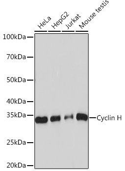
Western blot analysis of various lysates using Cyclin H Rabbit mAb (CAB4076) at 1:1000 dilution. Secondary antibody: HRP-conjugated Goat anti-Rabbit IgG (H+L) (AS014) at 1:10000 dilution. Lysates/proteins: 25μg per lane. Blocking buffer: 3% nonfat dry milk in TBST. Detection: ECL Basic Kit (AbGn00020). Exposure time: 60s.
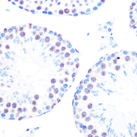
Immunohistochemistry analysis of paraffin-embedded Mouse testis using Cyclin H Rabbit mAb (CAB4076) at dilution of 1:100 (40x lens). Microwave antigen retrieval performed with 0.01M PBS Buffer (pH 7.2) prior to IHC staining.
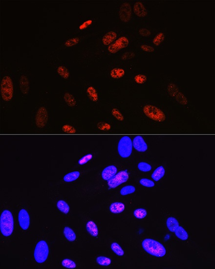
Immunofluorescence analysis of U-2 OS cells using Cyclin H Rabbit mAb (CAB4076) at dilution of 1:100 (40x lens). Secondary antibody: Cy3-conjugated Goat anti-Rabbit IgG (H+L) (AS007) at 1:500 dilution. Blue: DAPI for nuclear staining.


")