The CYP1A1 Antibody (CAB2159) is a high-quality antibody developed for reliable detection and analysis of target proteins. This antibody, raised in rabbits, exhibits high reactivity with human samples and is specifically validated for use in Western blot applications.Cytochrome P450 1A1 is known for its role in the activation and detoxification of environmental procarcinogens, making it a target for investigations in cancer research and toxicology. By binding to the cytochrome P450 1A1 protein, this antibody enables precise detection and analysis in a variety of cell types, facilitating studies on drug metabolism, environmental toxicity, and the development of novel therapeutic interventions.
This antibody is validated for use in WB, IF/ICC, ELISA applications and has demonstrated reactivity against Human, Mouse, Rat samples.
Product Name:
CYP1A1 Antibody
SKU:
CAB2159
Size:
20μL, 100μL
Reactivity:
Human, Mouse, Rat
Conjugate:
Unconjugated
Immunogen:
Recombinant protein (or fragment).This information is considered to be commercially sensitive.
This gene, CYP1A1, encodes a member of the cytochrome P450 superfamily of enzymes. The cytochrome P450 proteins are monooxygenases which catalyze many reactions involved in drug metabolism and synthesis of cholesterol, steroids and other lipids. This protein localizes to the endoplasmic reticulum and its expression is induced by some polycyclic aromatic hydrocarbons (PAHs), some of which are found in cigarette smoke. The enzyme's endogenous substrate is unknown; however, it is able to metabolize some PAHs to carcinogenic intermediates. The gene has been associated with lung cancer risk. A related family member, CYP1A2, is located approximately 25 kb away from CYP1A1 on chromosome 15. Alternative splicing results in multiple transcript variants encoding distinct isoforms.
Purification Method
Affinity purification
Gene ID
1543
RRID
AB_2764177
Buffer Information
Store at -20℃. Avoid freeze / thaw cycles. Buffer: PBS containing 50% glycerol, preserved with proclin300 or sodium azide, pH 7.3.
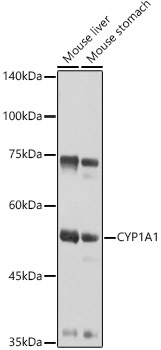
Western blot analysis of various lysates using CYP1A1 Rabbit pAb (CAB2159) at 1:1000 dilution. Secondary antibody: HRP-conjugated Goat anti-Rabbit IgG (H+L) (CABS014) at 1:10000 dilution. Lysates/proteins: 25μg per lane. Blocking buffer: 3% nonfat dry milk in TBST. Detection: ECL Basic Kit (AbGn00020). Exposure time: 3s.
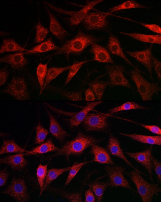
Immunofluorescence analysis of NIH/3T3 cells using CYP1A1 Rabbit pAb (CAB2159) at dilution of 1:100 (40x lens). Secondary antibody: Cy3-conjugated Goat anti-Rabbit IgG (H+L) (CABS007) at 1:500 dilution. Blue: DAPI for nuclear staining.