The CYR61 Antibody (CAB1111) is a high-quality antibody developed for reliable detection and analysis of target proteins. This antibody, produced in rabbits, demonstrates high specificity and sensitivity for detecting CYR61 in human samples, making it ideal for use in Western blot and immunohistochemistry applications.CYR61, also known as cysteine-rich protein 61, is involved in various physiological processes, including wound healing, tissue regeneration, and cancer progression. Its dysregulation has been implicated in the pathogenesis of multiple diseases, making it a promising target for therapeutic interventions.
This antibody is validated for use in WB, IF/ICC, ELISA applications and has demonstrated reactivity against Human, Mouse, Rat samples.
Product Name:
CYR61 Antibody
SKU:
CAB1111
Size:
20μL, 100μL
Reactivity:
Human, Mouse, Rat
Conjugate:
Unconjugated
Immunogen:
Synthetic peptide. This information is considered to be commercially sensitive.
Recommended starting concentration is 1 μg/mL. Please optimize the concentration based on your specific assay requirements.
Synonyms:
GIG1, CYR61, IGFBP10
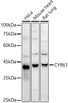
Positive Sample:
HeLa, Mouse heart, Rat lung
Cellular Localization:
Secreted.
Calculated MW:
42kDa
Observed MW:
41kDa
The secreted protein encoded by this gene is growth factor-inducible and promotes the adhesion of endothelial cells. The encoded protein interacts with several integrins and with heparan sulfate proteoglycan. This protein also plays a role in cell proliferation, differentiation, angiogenesis, apoptosis, and extracellular matrix formation.
Purification Method
Affinity purification
Gene ID
3491
RRID
AB_2758410
Buffer Information
Store at -20℃. Avoid freeze / thaw cycles. Buffer: PBS containing 50% glycerol, preserved with proclin300 or sodium azide, pH 7.3.
Western blot analysis of various lysates using CYR61 Rabbit pAb (CAB1111) at 1:1000 dilution. Secondary antibody: HRP-conjugated Goat anti-Rabbit IgG (H+L) (CABS014) at 1:10000 dilution. Lysates/proteins: 25μg per lane. Blocking buffer: 3% nonfat dry milk in TBST. Detection: ECL Basic Kit (AbGn00020). Exposure time: 30s.
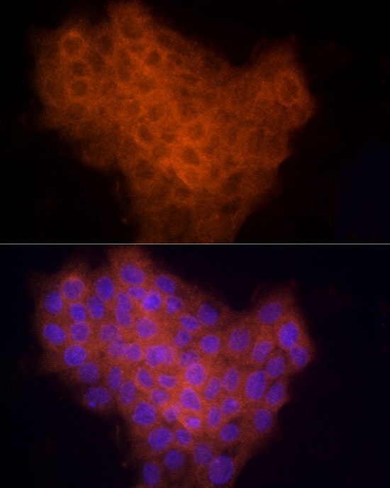
Immunofluorescence analysis of A431 cells using CYR61 Rabbit pAb (CAB1111) at dilution of 1:200 (40x lens). Secondary antibody: Cy3-conjugated Goat anti-Rabbit IgG (H+L) (CABS007) at 1:500 dilution. Blue: DAPI for nuclear staining.