The Cytokeratin 7 (KRT7) Monoclonal Antibody (CAB4765) is a high-quality antibody developed for reliable detection and analysis of target proteins. This high-quality antibody, produced in rabbits, is specifically designed for use in immunohistochemistry (IHC) applications, where it shows strong reactivity with human samples.Cytokeratin 7 is a marker commonly used in cancer research, as its expression pattern can provide valuable information about tumor origin and differentiation. The CAB4765 antibody binds specifically to cytokeratin 7, allowing for precise detection and localization of this protein in a variety of tissue samples. Its reliability and sensitivity make it an essential reagent for studies focusing on epithelial cell biology, tumor biomarker analysis, and diagnostic pathology.
This antibody is validated for use in WB, IHC-P, ELISA, IF-P applications and has demonstrated reactivity against Human, Mouse, Rat samples.
Product Name:
Cytokeratin 7 (KRT7) Monoclonal Antibody
SKU:
CAB4765
Size:
20μL, 100μL
Reactivity:
Human, Mouse, Rat
Clone Number:
ARC1267
Conjugate:
Unconjugated
Immunogen:
Synthetic peptide. This information is considered to be commercially sensitive.
Recommended starting concentration is 1 μg/mL. Please optimize the concentration based on your specific assay requirements.
Synonyms:
K7, CK7, SCL, K2C7, Cytokeratin 7 (KRT7)
Positive Sample:
HeLa, A-549, Mouse lung, Mouse kidney, Rat kidney
Cellular Localization:
Cytoplasm.
Calculated MW:
51kDa
Observed MW:
55kDa
The protein encoded by this gene is a member of the keratin gene family. The type II cytokeratins consist of basic or neutral proteins which are arranged in pairs of heterotypic keratin chains coexpressed during differentiation of simple and stratified epithelial tissues. This type II cytokeratin is specifically expressed in the simple epithelia lining the cavities of the internal organs and in the gland ducts and blood vessels. The genes encoding the type II cytokeratins are clustered in a region of chromosome 12q12-q13. Alternative splicing may result in several transcript variants; however, not all variants have been fully described.
Purification Method
Affinity purification
Gene ID
3855
RRID
AB_2863343
Buffer Information
Store at -20℃. Avoid freeze / thaw cycles. Buffer: PBS containing 50% glycerol and 0.05% BSA, preserved with proclin300 or sodium azide, pH 7.3.
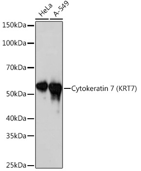
Western blot analysis of various lysates using Cytokeratin 7 (KRT7) (KRT7) Rabbit mAb (CAB4765) at 1:1000 dilution. Secondary antibody: HRP-conjugated Goat anti-Rabbit IgG (H+L) (CABS014) at 1:10000 dilution. Lysates/proteins: 25μg per lane. Blocking buffer: 3% nonfat dry milk in TBST. Detection: ECL Basic Kit (AbGn00020). Exposure time: 10s.
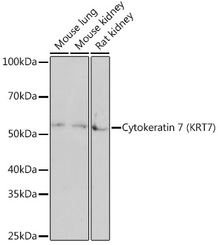
Western blot analysis of various lysates using Cytokeratin 7 (KRT7) (KRT7) Rabbit mAb (CAB4765) at 1:1000 dilution. Secondary antibody: HRP-conjugated Goat anti-Rabbit IgG (H+L) (CABS014) at 1:10000 dilution. Lysates/proteins: 25μg per lane. Blocking buffer: 3% nonfat dry milk in TBST. Detection: ECL Basic Kit (AbGn00020). Exposure time: 3min.
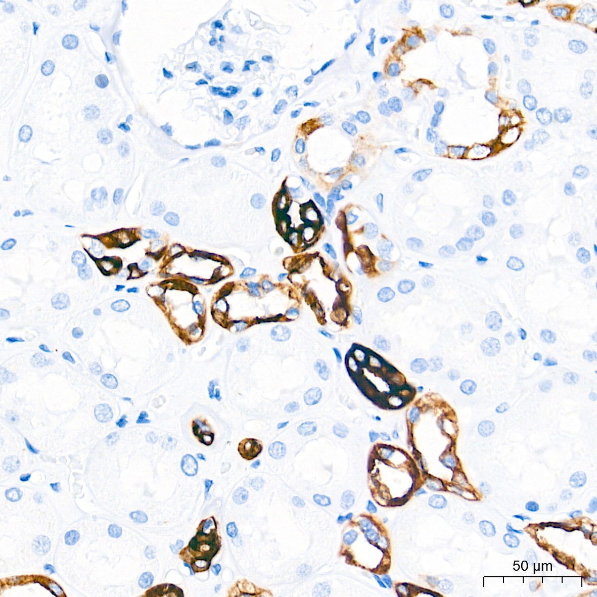
Immunohistochemistry analysis of paraffin-embedded Human kidney tissue using Cytokeratin 7 (KRT7) Rabbit mAb (CAB4765) at a dilution of 1:1000 (40x lens). High pressure antigen retrieval performed with 0.01M Tris-EDTA Buffer (pH 9.0) prior to IHC staining.
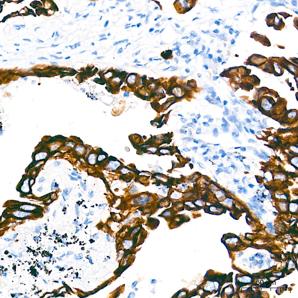
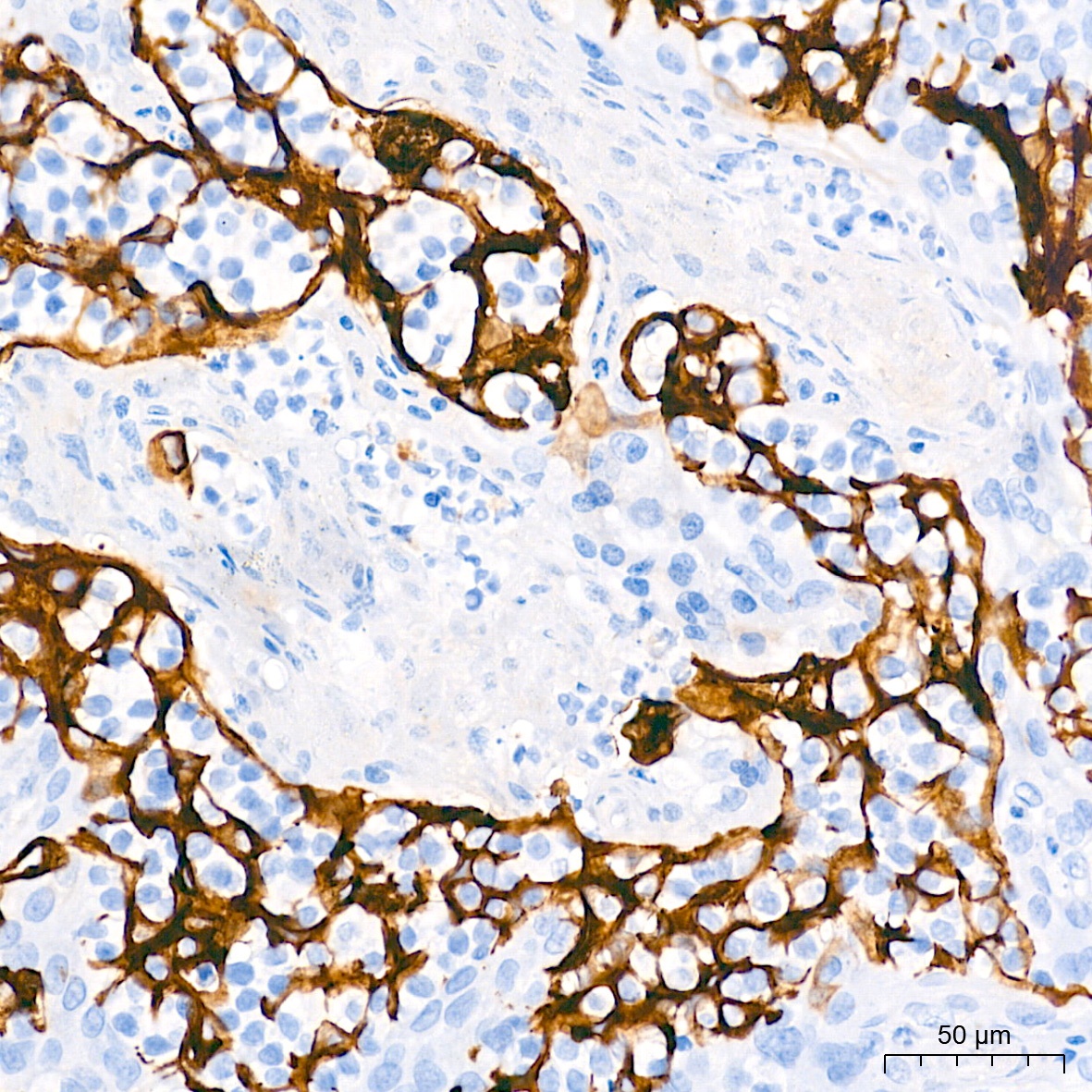
Immunohistochemistry analysis of paraffin-embedded Human lung cancer tissue using Cytokeratin 7 (KRT7) Rabbit mAb (CAB4765) at a dilution of 1:1000 (40x lens). High pressure antigen retrieval performed with 0.01M Tris-EDTA Buffer (pH 9.0) prior to IHC staining.
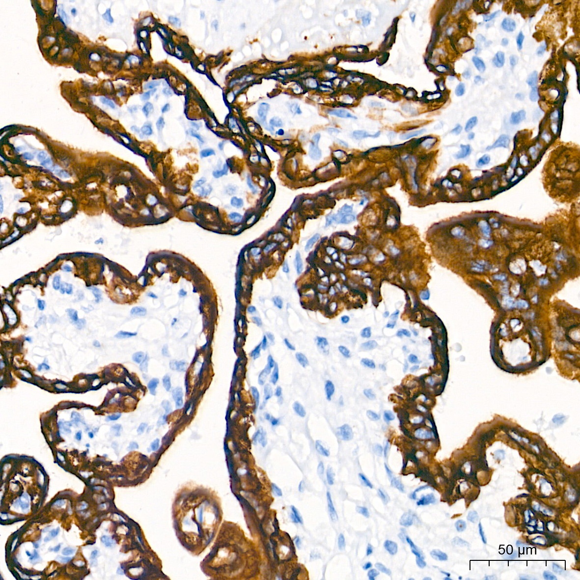
Immunohistochemistry analysis of paraffin-embedded Human placenta tissue using Cytokeratin 7 (KRT7) Rabbit mAb (CAB4765) at a dilution of 1:1000 (40x lens). High pressure antigen retrieval performed with 0.01M Tris-EDTA Buffer (pH 9.0) prior to IHC staining.
Immunohistochemistry analysis of paraffin-embedded Human tonsil tissue using Cytokeratin 7 (KRT7) Rabbit mAb (CAB4765) at a dilution of 1:1000 (40x lens). High pressure antigen retrieval performed with 0.01M Tris-EDTA Buffer (pH 9.0) prior to IHC staining.