The DCD Antibody (CAB7280) is a high-quality antibody developed for reliable detection and analysis of target proteins. This antibody, produced in rabbits, exhibits high reactivity with human samples and is optimized for Western blot applications. By specifically binding to dermcidin, researchers can accurately detect and analyze this important protein in a variety of cell types, making it an invaluable tool for studies in dermatology, microbiology, and infectious disease research.
This antibody is validated for use in WB, ELISA applications and has demonstrated reactivity against Human, Mouse samples.
Product Name:
DCD Antibody
SKU:
CAB7280
Size:
20μL, 100μL
Reactivity:
Human, Mouse
Conjugate:
Unconjugated
Immunogen:
Recombinant protein (or fragment).This information is considered to be commercially sensitive.
Recommended starting concentration is 1 μg/mL. Please optimize the concentration based on your specific assay requirements.
Synonyms:
PIF, AIDD, DSEP, HCAP, DCD-1, DCD
Positive Sample:
HeLa, 293T
Cellular Localization:
Secreted.
Calculated MW:
11kDa
Observed MW:
18kDa
This antimicrobial gene encodes a secreted protein that is subsequently processed into mature peptides of distinct biological activities. The C-terminal peptide is constitutively expressed in sweat and has antibacterial and antifungal activities. The N-terminal peptide, also known as diffusible survival evasion peptide, promotes neural cell survival under conditions of severe oxidative stress. A glycosylated form of the N-terminal peptide may be associated with cachexia (muscle wasting) in cancer patients. Alternative splicing results in multiple transcript variants encoding different isoforms.
Purification Method
Affinity purification
Gene ID
117159
RRID
AB_2767821
Buffer Information
Store at -20℃. Avoid freeze / thaw cycles. Buffer: PBS containing 50% glycerol, preserved with proclin300 or sodium azide, pH 7.3.
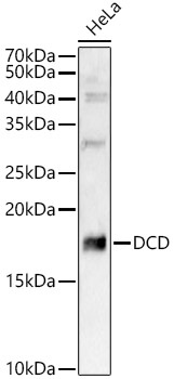
Western blot analysis of lysates from HeLa cells, using DCD Rabbit pAb (CAB7280) at 1:8000 dilution. Secondary antibody: HRP-conjugated Goat anti-Rabbit IgG (H+L) (CABS014) at 1:10000 dilution. Lysates/proteins: 25μg per lane. Blocking buffer: 3% nonfat dry milk in TBST. Detection: ECL Basic Kit (AbGn00020). Exposure time: 90s.
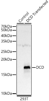
Western blot analysis of lysates from 293T cells, using DCD Rabbit pAb (CAB7280) at 1:8000 dilution. Secondary antibody: HRP-conjugated Goat anti-Rabbit IgG (H+L) (CABS014) at 1:10000 dilution. Lysates/proteins: 25μg per lane. Blocking buffer: 3% nonfat dry milk in TBST. Detection: ECL Basic Kit (AbGn00020). Exposure time: 90s.