The DCX Antibody (CAB1134) is a high-quality antibody developed for reliable detection and analysis of target proteins. This antibody, produced in rabbits, shows strong reactivity with human samples and is validated for use in Western blot applications. By binding specifically to the DCX protein, this antibody allows for accurate detection and analysis in various cell types, making it ideal for studies in neuroscience and neurodevelopmental disorders.DCX, also known as doublecortin, is crucial for proper neuronal migration and organization in the developing brain. Mutations in the DCX gene have been linked to disorders like lissencephaly and subcortical band heterotopia, highlighting its importance in brain development.
This antibody is validated for use in WB, IF/ICC, ELISA applications and has demonstrated reactivity against Human, Mouse, Rat samples.
Product Name:
DCX Antibody
SKU:
CAB1134
Size:
20μL, 100μL
Reactivity:
Human, Mouse, Rat
Conjugate:
Unconjugated
Immunogen:
Recombinant protein (or fragment).This information is considered to be commercially sensitive.
Recommended starting concentration is 1 μg/mL. Please optimize the concentration based on your specific assay requirements.
Synonyms:
DC, DBCN, LISX, SCLH, XLIS, Doublecortin
Positive Sample:
SH-SY5Y, Mouse spinal cord
Cellular Localization:
Cell Projection, Cytoplasm.
Calculated MW:
41kDa
Observed MW:
45kDa
This gene encodes a member of the doublecortin family. The protein encoded by this gene is a cytoplasmic protein and contains two doublecortin domains, which bind microtubules. In the developing cortex, cortical neurons must migrate over long distances to reach the site of their final differentiation. The encoded protein appears to direct neuronal migration by regulating the organization and stability of microtubules. In addition, the encoded protein interacts with LIS1, the regulatory gamma subunit of platelet activating factor acetylhydrolase, and this interaction is important to proper microtubule function in the developing cortex. Mutations in this gene cause abnormal migration of neurons during development and disrupt the layering of the cortex, leading to epilepsy, cognitive disability, subcortical band heterotopia ("double cortex" syndrome) in females and lissencephaly ("smooth brain" syndrome) in males. Multiple transcript variants encoding different isoforms have been found for this gene.
Purification Method
Affinity purification
Gene ID
1641
RRID
AB_2758515
Buffer Information
Store at -20℃. Avoid freeze / thaw cycles. Buffer: PBS containing 50% glycerol, preserved with proclin300 or sodium azide, pH 7.3.
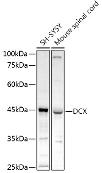
Western blot analysis of various lysates using Doublecortin Rabbit pAb (CAB1134) at 1:1000 dilution. Secondary antibody: HRP-conjugated Goat anti-Rabbit IgG (H+L) (CABS014) at 1:10000 dilution. Lysates/proteins: 25μg per lane. Blocking buffer: 3% nonfat dry milk in TBST. Detection: ECL Enhanced Kit (AbGn00021). Exposure time: 180s.
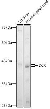
Western blot analysis of various lysates using Doublecortin Rabbit pAb (CAB1134) at 1:1000 dilution. Secondary antibody: HRP-conjugated Goat anti-Rabbit IgG (H+L) (CABS014) at 1:10000 dilution. Lysates/proteins: 25μg per lane. Blocking buffer: 3% nonfat dry milk in TBST. Detection: ECL Enhanced Kit (AbGn00021). Exposure time: 180s.
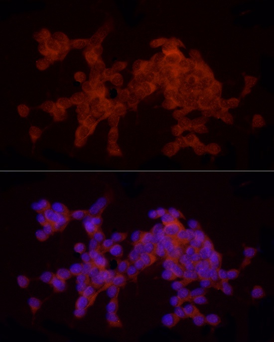
Immunofluorescence analysis of SH-5Y5Y cells using Doublecortin Rabbit pAb (CAB1134) at dilution of 1:20 (40x lens). Secondary antibody: Cy3-conjugated Goat anti-Rabbit IgG (H+L) (CABS007) at 1:500 dilution. Blue: DAPI for nuclear staining.
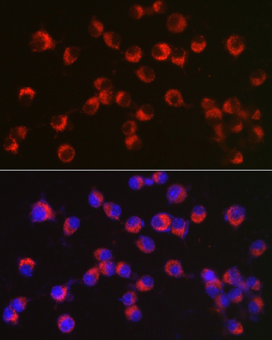
Immunofluorescence analysis of Neuro-2a cells using Doublecortin Rabbit pAb (CAB1134) at dilution of 1:100 (40x lens). Secondary antibody: Cy3-conjugated Goat anti-Rabbit IgG (H+L) (CABS007) at 1:500 dilution. Blue: DAPI for nuclear staining.