The DDB1 Antibody (CAB2896) is a high-quality antibody developed for reliable detection and analysis of target proteins. The protein encoded by this gene is the large subunit (p127) of the heterodimeric DNA damage-binding (DDB) complex while another protein (p48) forms the small subunit. This protein complex functions in nucleotide-excision repair and binds to DNA following UV damage. Defective activity of this complex causes the repair defect in patients with xeroderma pigmentosum complementation group E (XPE) - an autosomal recessive disorder characterized by photosensitivity and early onset of carcinomas. However, it remains for mutation analysis to demonstrate whether the defect in XPE patients is in this gene or the gene encoding the small subunit. In addition, Best vitelliform mascular dystrophy is mapped to the same region as this gene on 11q, but no sequence alternations of this gene are demonstrated in Best disease patients. The protein encoded by this gene also functions as an adaptor molecule for the cullin 4 (CUL4) ubiquitin E3 ligase complex by facilitating the binding of substrates to this complex and the ubiquitination of proteins. RRID AB_2764716 Gene ID 1642 Swiss Prot Synonym XPE; DDBA; XAP1; XPCE; XPE-BF; UV-DDB1; WHIKERS; DDB1
This antibody is validated for use in WB, IHC-P, IF/ICC, ELISA applications and has demonstrated reactivity against Human, Mouse, Rat samples.
Product Name:
DDB1 Antibody
SKU:
CAB2896
Size:
100μL, 20μL
Reactivity:
Human, Mouse, Rat
Clone Number:
-
Conjugate:
Unconjugated
Immunogen:
Synthetic peptide. This information is considered to be commercially sensitive.
Tested Applications:
WBIHC-PIF/ICCELISA
Recommended Dilution:
WB
1:500 - 1:1000
IHC-P
1:50 - 1:100
IF
/
ICC
1:50 - 1:100
ELISA
Recommended starting concentration is 1 μg/mL. Please optimize the concentration based on your specific assay requirements.
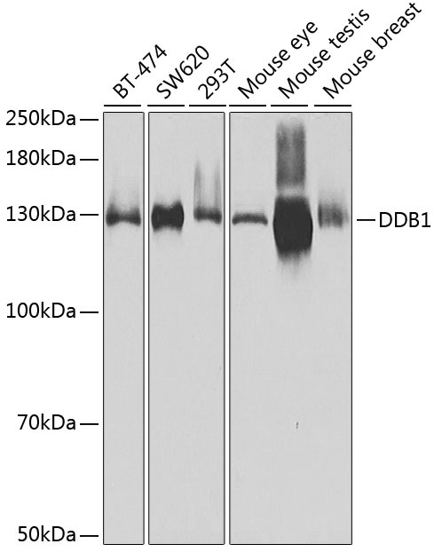
BT-474, SW620, 293T, Mouse eye, Mouse testis, Mouse breast
Cellular Localization:
Cytoplasm, Nucleus.
Calculated MW:
127kDa
Observed MW:
127kDa
The protein encoded by this gene is the large subunit (p127) of the heterodimeric DNA damage-binding (DDB) complex while another protein (p48) forms the small subunit. This protein complex functions in nucleotide-excision repair and binds to DNA following UV damage. Defective activity of this complex causes the repair defect in patients with xeroderma pigmentosum complementation group E (XPE) - an autosomal recessive disorder characterized by photosensitivity and early onset of carcinomas. However, it remains for mutation analysis to demonstrate whether the defect in XPE patients is in this gene or the gene encoding the small subunit. In addition, Best vitelliform mascular dystrophy is mapped to the same region as this gene on 11q, but no sequence alternations of this gene are demonstrated in Best disease patients. The protein encoded by this gene also functions as an adaptor molecule for the cullin 4 (CUL4) ubiquitin E3 ligase complex by facilitating the binding of substrates to this complex and the ubiquitination of proteins. RRID AB_2764716 Gene ID 1642 Swiss Prot Synonym XPE; DDBA; XAP1; XPCE; XPE-BF; UV-DDB1; WHIKERS; DDB1
Purification Method:
Affinity purification
Gene ID:
1642
RRID:
AB_2764716
Buffer Information:
Store at -20℃. Avoid freeze / thaw cycles. Buffer: PBS with 0.09% Sodium azide,50% glycerol,pH7.3.
Western blot analysis of various lysates using DDB1 Rabbit pAb (CAB2896) at 1:500 dilution. Secondary antibody: HRP-conjugated Goat anti-Rabbit IgG (H+L) (AS014) at 1:10000 dilution. Lysates/proteins: 25μg per lane. Blocking buffer: 3% nonfat dry milk in TBST. Detection: ECL Basic Kit (AbGn00020). Exposure time: 30s.
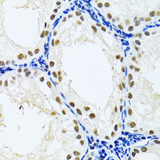
Immunohistochemistry analysis of paraffin-embedded Rat testis using DDB1 Rabbit pAb (CAB2896) at dilution of 1:100 (40x lens). Microwave antigen retrieval performed with 0.01M PBS Buffer (pH 7.2) prior to IHC staining.
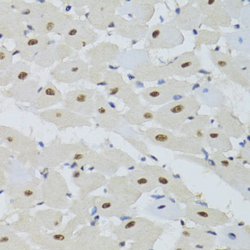
Immunohistochemistry analysis of paraffin-embedded Mouse heart using DDB1 Rabbit pAb (CAB2896) at dilution of 1:100 (40x lens). Microwave antigen retrieval performed with 0.01M PBS Buffer (pH 7.2) prior to IHC staining.
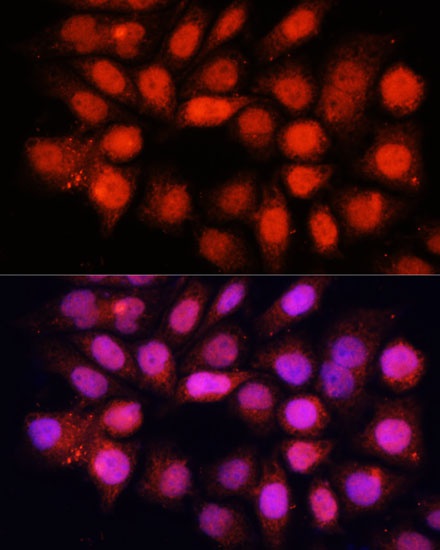
Immunofluorescence analysis of HeLa cells using DDB1 Rabbit pAb (CAB2896) at dilution of 1:100 (40x lens). Secondary antibody: Cy3-conjugated Goat anti-Rabbit IgG (H+L) (AS007) at 1:500 dilution. Blue: DAPI for nuclear staining.