The DDIT3/CHOP Antibody (CAB11346) is a high-quality antibody developed for reliable detection and analysis of target proteins. This antibody, produced in rabbits, exhibits high reactivity with human samples and has been validated for use in Western blot applications. By specifically binding to the DDIT3 protein, researchers can easily detect and analyze its expression in a variety of cell types, making it an ideal choice for studies in molecular biology and cancer research.
This antibody is validated for use in WB, IHC-P, IP, ELISA applications and has demonstrated reactivity against Mouse, Rat samples.
Product Name:
DDIT3/CHOP Antibody
SKU:
CAB11346
Size:
20μL, 100μL
Reactivity:
Mouse, Rat
Conjugate:
Unconjugated
Immunogen:
Synthetic peptide. This information is considered to be commercially sensitive.
This gene encodes a member of the CCAAT/enhancer-binding protein (C/EBP) family of transcription factors. The protein functions as a dominant-negative inhibitor by forming heterodimers with other C/EBP members, such as C/EBP and LAP (liver activator protein), and preventing their DNA binding activity. The protein is implicated in adipogenesis and erythropoiesis, is activated by endoplasmic reticulum stress, and promotes apoptosis. Fusion of this gene and FUS on chromosome 16 or EWSR1 on chromosome 22 induced by translocation generates chimeric proteins in myxoid liposarcomas or Ewing sarcoma. Multiple alternatively spliced transcript variants encoding two isoforms with different length have been identified.
Purification Method
Affinity purification
Gene ID
1649
RRID
AB_2758518
Buffer Information
Store at -20℃. Avoid freeze / thaw cycles. Buffer: PBS containing 50% glycerol, preserved with proclin300 or sodium azide, pH 7.3.
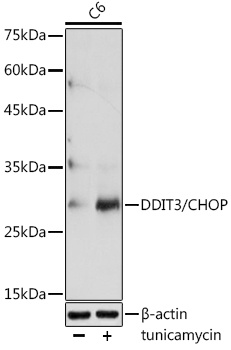
Western blot analysis of lysates from C6 cells, using DDIT3/CHOP Rabbit pAb (CAB11346) at 1:1000 dilution. C6 cells were treated with tunicamycin (2 μg/ml) for 8 hours. Secondary antibody: HRP-conjugated Goat anti-Rabbit IgG (H+L) (CABS014) at 1:10000 dilution. Lysates/proteins: 25μg per lane. Blocking buffer: 3% nonfat dry milk in TBST. Detection: ECL Basic Kit (AbGn00020). Exposure time: 90s.
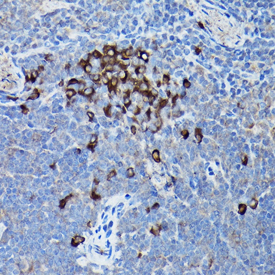
Immunohistochemistry analysis of paraffin-embedded Mouse spleen using DDIT3/CHOP Rabbit pAb (CAB11346) at dilution of 1:200 (40x lens). High pressure antigen retrieval performed with 0.01M Citrate buffer (pH 6.0) prior to IHC staining.
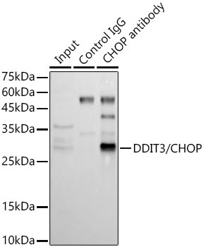
Immunoprecipitation analysis of 300 μg extracts of C2C12 tunicamycin cells using 3 μg DDIT3/CHOP antibody (CAB11346). Western blot was performed from the immunoprecipitate using DDIT3/CHOP antibody (CAB11346) at a dilution of 1:500.