The DAB1 Antibody (CAB10349) is a high-quality antibody developed for reliable detection and analysis of target proteins. The laminar organization of multiple neuronal types in the cerebral cortex is required for normal cognitive function. In mice, the disabled-1 gene plays a central role in brain development, directing the migration of cortical neurons past previously formed neurons to reach their proper layer. This gene is similar to disabled-1, and the protein encoded by this gene is thought to be a signal transducer that interacts with protein kinase pathways to regulate neuronal positioning in the developing brain.
This antibody is validated for use in WB, IF/ICC, ELISA applications and has demonstrated reactivity against Human, Mouse, Rat samples.
Product Name:
DAB1 Antibody
SKU:
CAB10349
Size:
100μL, 20μL
Reactivity:
Human, Mouse, Rat
Conjugate:
Unconjugated
Immunogen:
Recombinant protein (or fragment).This information is considered to be commercially sensitive.
Tested Applications:
WBIF/ICCELISA
Recommended Dilution:
WB
1:100 - 1:500
IF/ICC
1:50 - 1:200
ELISA
Recommended starting concentration is 1 μg/mL. Please optimize the concentration based on your specific assay requirements.
Synonyms:
SCA37, DAB1
Positive Sample:
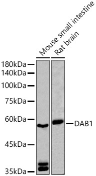
Mouse small intestine, Rat brain
Cellular Localization:
Cytoplasm, Cytosol, Perinuclear Region Of Cytoplasm.
Calculated MW:
64kDa
Observed MW:
57kDa/59kDa
The laminar organization of multiple neuronal types in the cerebral cortex is required for normal cognitive function. In mice, the disabled-1 gene plays a central role in brain development, directing the migration of cortical neurons past previously formed neurons to reach their proper layer. This gene is similar to disabled-1, and the protein encoded by this gene is thought to be a signal transducer that interacts with protein kinase pathways to regulate neuronal positioning in the developing brain.
Purification Method
Affinity purification
Gene ID
1600
RRID
AB_2757894
Buffer Information
Store at -20℃. Avoid freeze / thaw cycles. Buffer: PBS containing 50% glycerol, preserved with proclin300 or sodium azide, pH 7.3.
Western blot analysis of various lysates using DAB1 Rabbit pAb (CAB10349) at 1:500 dilution. Secondary antibody: HRP-conjugated Goat anti-Rabbit IgG (H+L) (AS014) at 1:10000 dilution. Lysates/proteins: 25μg per lane. Blocking buffer: 3% nonfat dry milk in TBST. Detection: ECL Basic Kit (AbGn00020). Exposure time: 90s.
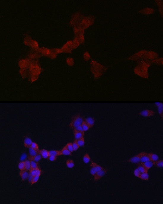
Immunofluorescence analysis of SH-SY5Y cells using DAB1 Rabbit pAb (CAB10349) at dilution of 1:200 (40x lens). Secondary antibody: Cy3-conjugated Goat anti-Rabbit IgG (H+L) (AS007) at 1:500 dilution. Blue: DAPI for nuclear staining.