The DKC1 Monoclonal Antibody (CAB4407) is a high-quality antibody developed for reliable detection and analysis of target proteins. This antibody, produced in rabbits, is highly specific for DKC1 and has been validated for use in various applications, including Western blot and immunohistochemistry.DKC1 is a multifunctional protein that plays a critical role in cell growth and division, making it a key target for investigation in cancer and other diseases. Dysregulation of DKC1 has been implicated in various cancers, making this antibody valuable for studies aiming to understand the molecular mechanisms underlying these diseases.
This antibody is validated for use in WB, IHC-P, ELISA applications and has demonstrated reactivity against Human, Mouse, Rat samples.
Product Name:
DKC1 Monoclonal Antibody
SKU:
CAB4407
Size:
20μL, 100μL
Reactivity:
Human, Mouse, Rat
Clone Number:
ARC1063
Conjugate:
Unconjugated
Immunogen:
Synthetic peptide. This information is considered to be commercially sensitive.
Recommended starting concentration is 1 μg/mL. Please optimize the concentration based on your specific assay requirements.
Synonyms:
DKC, CBF5, DKCX, NAP57, NOLA4, XAP101, DKC1
Positive Sample:
PC-3, Mouse brain, Mouse heart, Rat liver
Cellular Localization:
Cajal Body, Cytoplasm, Nucleus, Nucleolus.
Calculated MW:
58kDa
Observed MW:
60kDa
This gene functions in two distinct complexes. It plays an active role in telomerase stabilization and maintenance, as well as recognition of snoRNAs containing H/ACA sequences which provides stability during biogenesis and assembly into H/ACA small nucleolar RNA ribonucleoproteins (snoRNPs). This gene is highly conserved and widely expressed, and may play additional roles in nucleo-cytoplasmic shuttling, DNA damage response, and cell adhesion. Mutations have been associated with X-linked dyskeratosis congenita. Alternative splicing results in multiple transcript variants.
Purification Method
Affinity purification
Gene ID
1736
RRID
AB_2863266
Buffer Information
Store at -20℃. Avoid freeze / thaw cycles. Buffer: PBS containing 50% glycerol and 0.05% BSA, preserved with proclin300 or sodium azide, pH 7.3.
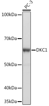
Western blot analysis of lysates from PC-3 cells, using DKC1 Rabbit mAb (CAB4407) at 1:1000 dilution. Secondary antibody: HRP-conjugated Goat anti-Rabbit IgG (H+L) (CABS014) at 1:10000 dilution. Lysates/proteins: 25μg per lane. Blocking buffer: 3% nonfat dry milk in TBST. Detection: ECL Basic Kit (AbGn00020). Exposure time: 1s.
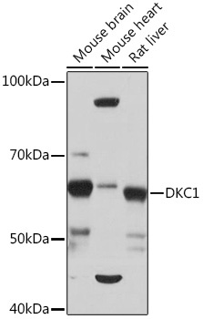
Western blot analysis of various lysates using DKC1 Rabbit mAb (CAB4407) at 1:1000 dilution. Secondary antibody: HRP-conjugated Goat anti-Rabbit IgG (H+L) (CABS014) at 1:10000 dilution. Lysates/proteins: 25μg per lane. Blocking buffer: 3% nonfat dry milk in TBST. Detection: ECL Basic Kit (AbGn00020). Exposure time: 10s.
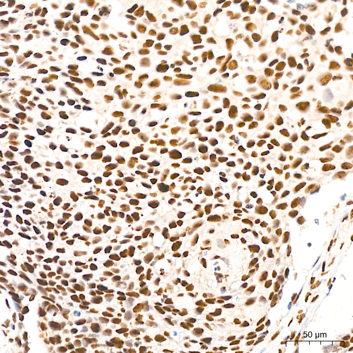
Immunohistochemistry analysis of paraffin-embedded Human cervix cancer tissue using DKC1 Rabbit mAb (CAB4407) at a dilution of 1:200 (40x lens). High pressure antigen retrieval was performed with 0.01 M citrate buffer (pH 6.0) prior to IHC staining.
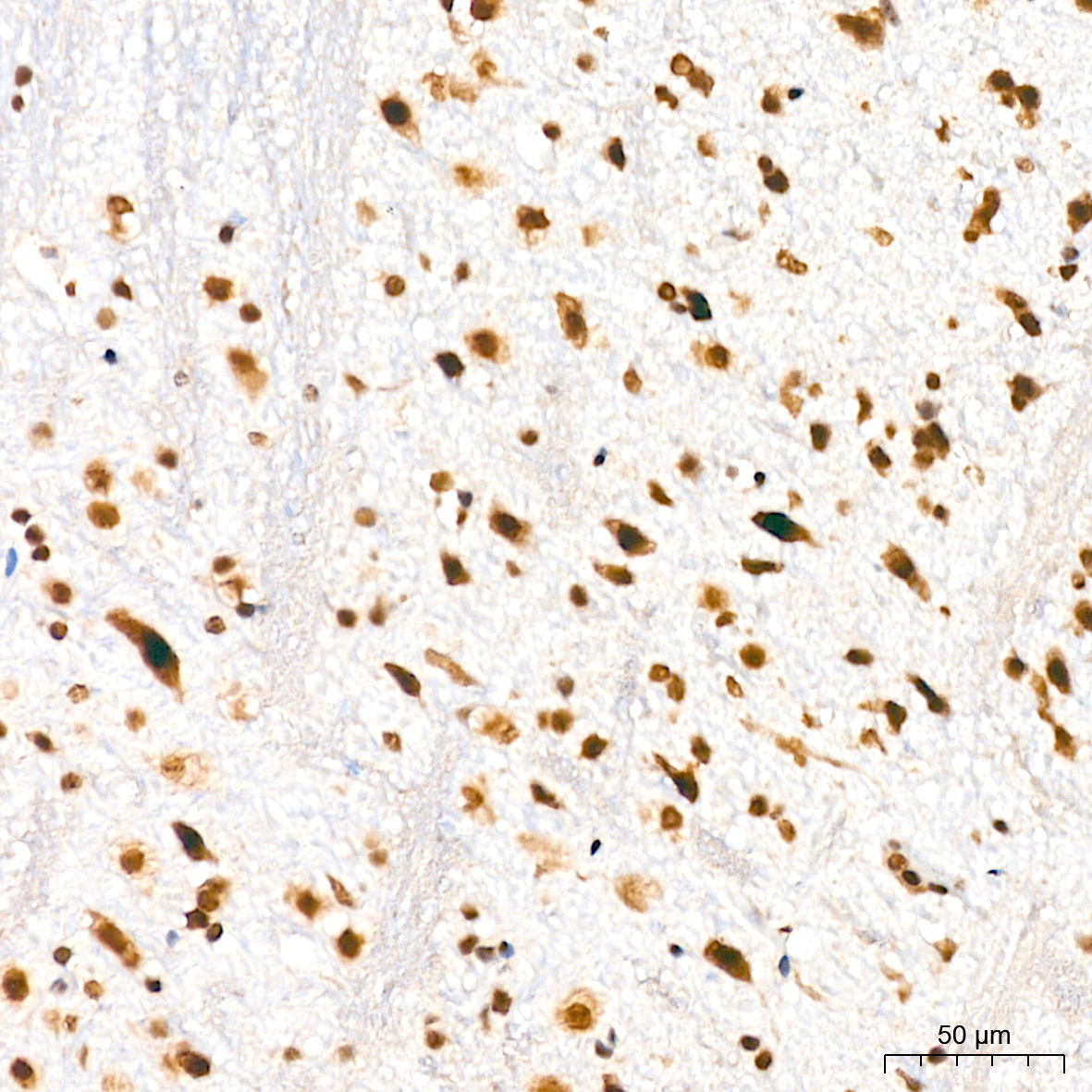
Immunohistochemistry analysis of paraffin-embedded Mouse brain tissue using DKC1 Rabbit mAb (CAB4407) at a dilution of 1:200 (40x lens). High pressure antigen retrieval was performed with 0.01 M citrate buffer (pH 6.0) prior to IHC staining.
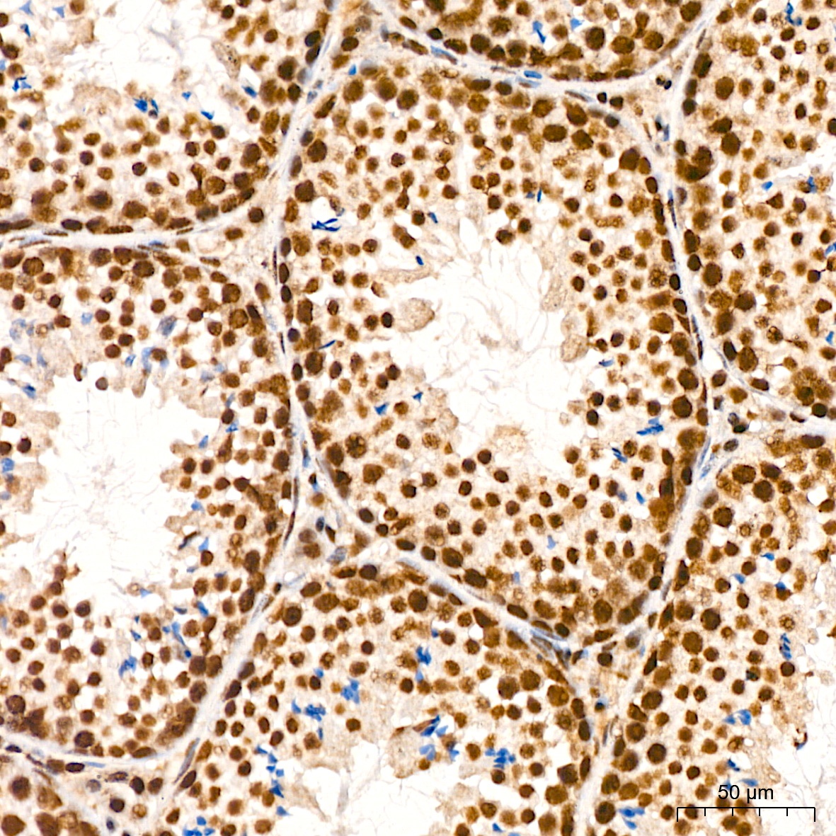
Immunohistochemistry analysis of paraffin-embedded Mouse testis tissue using DKC1 Rabbit mAb (CAB4407) at a dilution of 1:200 (40x lens). High pressure antigen retrieval was performed with 0.01 M citrate buffer (pH 6.0) prior to IHC staining.
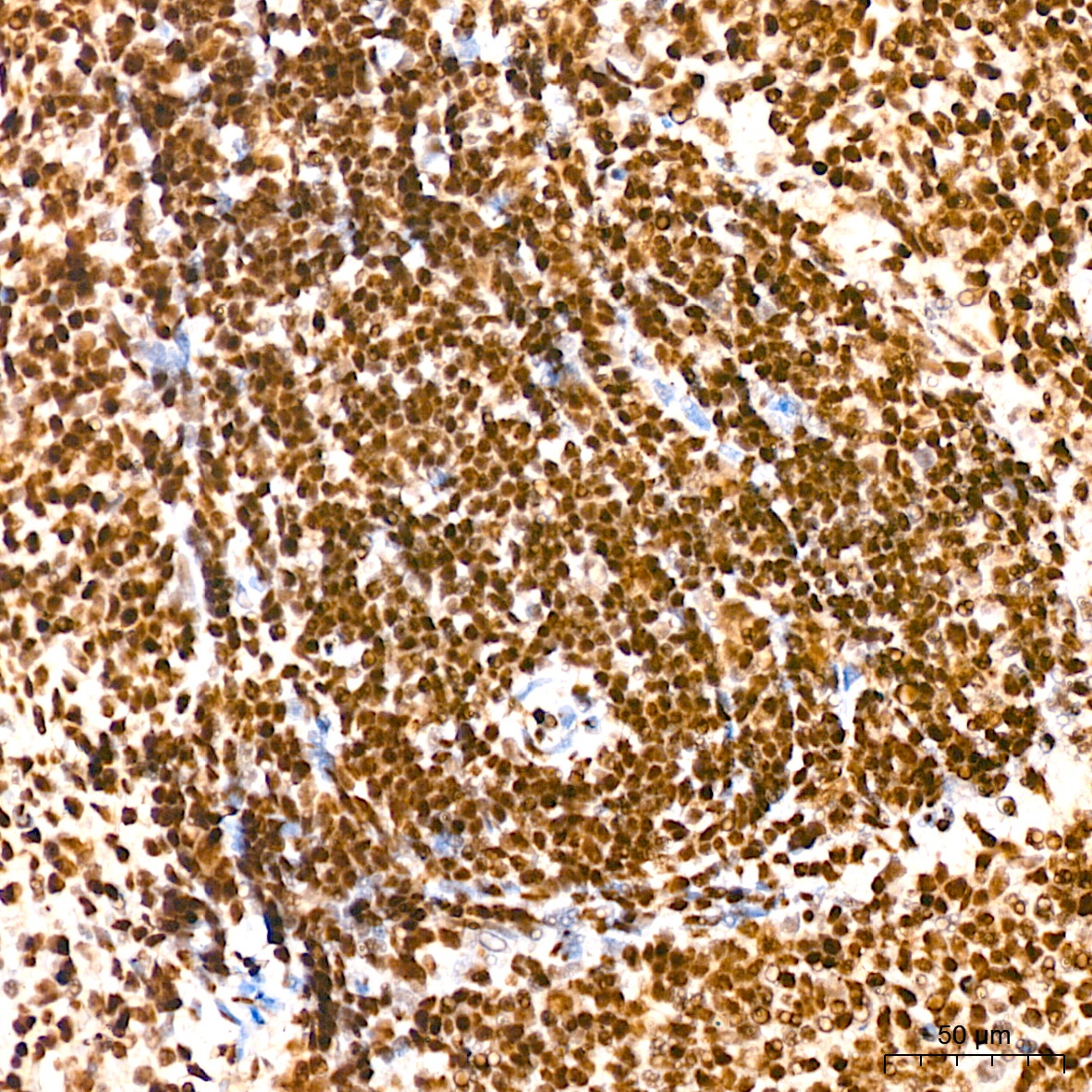
Immunohistochemistry analysis of paraffin-embedded Rat spleen tissue using DKC1 Rabbit mAb (CAB4407) at a dilution of 1:200 (40x lens). High pressure antigen retrieval was performed with 0.01 M citrate buffer (pH 6.0) prior to IHC staining.