The DRP1 Antibody (CAB16661) is a high-quality antibody developed for reliable detection and analysis of target proteins. This antibody, generated in rabbits, is highly specific to human samples and has been validated for use in Western blot applications. By binding to DNM1L, this antibody allows for the detection and analysis of this protein in various cell types, making it ideal for investigations in mitochondrial biology and diseases related to dysfunction in mitochondrial dynamics.DNM1L, also known as dynamin-1-like protein, plays a crucial role in mitochondrial fission, a process essential for maintaining mitochondrial function and cellular homeostasis.
This antibody is validated for use in WB, IHC-P, IF/ICC, ELISA applications and has demonstrated reactivity against Human, Mouse, Rat samples.
Product Name:
DRP1 Antibody
SKU:
CAB16661
Size:
20μL, 100μL
Reactivity:
Human, Mouse, Rat
Conjugate:
Unconjugated
Immunogen:
Recombinant protein (or fragment).This information is considered to be commercially sensitive.
This gene encodes a member of the dynamin superfamily of GTPases. The encoded protein mediates mitochondrial and peroxisomal division, and is involved in developmentally regulated apoptosis and programmed necrosis. Dysfunction of this gene is implicated in several neurological disorders, including Alzheimer's disease. Mutations in this gene are associated with the autosomal dominant disorder, encephalopathy, lethal, due to defective mitochondrial and peroxisomal fission (EMPF). Alternative splicing results in multiple transcript variants encoding different isoforms.
Purification Method
Affinity purification
Gene ID
10059
RRID
AB_2769197
Buffer Information
Store at -20℃. Avoid freeze / thaw cycles. Buffer: PBS containing 50% glycerol, preserved with proclin300 or sodium azide, pH 7.3.
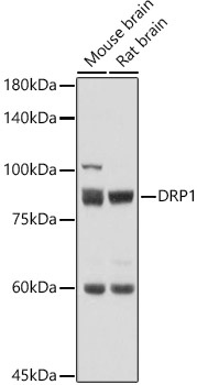
Western blot analysis of various lysates using DRP1 Rabbit pAb (CAB16661) at 1:500 dilution. Secondary antibody: HRP-conjugated Goat anti-Rabbit IgG (H+L) (CABS014) at 1:10000 dilution. Lysates/proteins: 25μg per lane. Blocking buffer: 3% nonfat dry milk in TBST. Detection: ECL Basic Kit (AbGn00020). Exposure time: 1s.
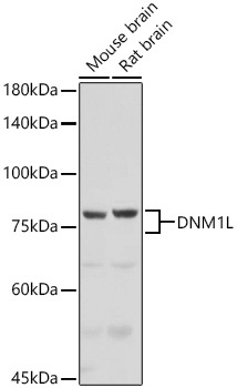
Western blot analysis of various lysates using DRP1 Rabbit pAb (CAB16661) at 1:1000 dilution incubated overnight at 4℃. Secondary antibody: HRP-conjugated Goat anti-Rabbit IgG (H+L) (CABS014) at 1:10000 dilution. Lysates/proteins: 25 μg per lane. Blocking buffer: 3% nonfat dry milk in TBST. Detection: ECL Basic Kit (AbGn00020). Exposure time: 3s.
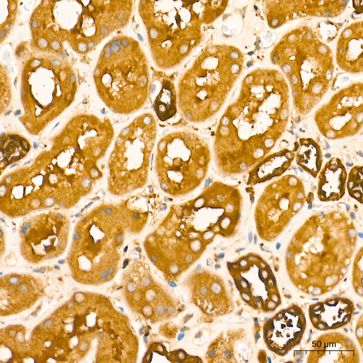
Immunohistochemistry analysis of paraffin-embedded Human kidney tissue using DRP1 Rabbit pAb (CAB16661) at a dilution of 1:300 (40x lens). High pressure antigen retrieval performed with 0.01M Tris-EDTA Buffer (pH 9.0) prior to IHC staining.
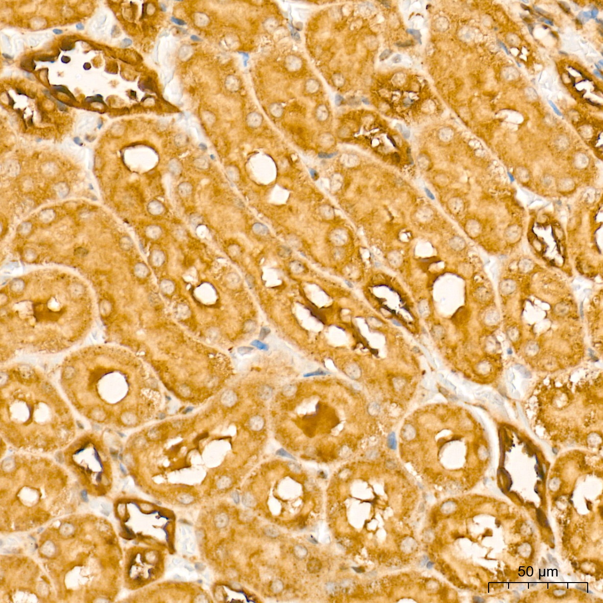
Immunohistochemistry analysis of paraffin-embedded rat kidney tissue using DRP1 Rabbit pAb (CAB16661) at a dilution of 1:300 (40x lens). High pressure antigen retrieval performed with 0.01M Tris-EDTA Buffer (pH 9.0) prior to IHC staining.